BOVINE IMAGES

How to use these images


IMPORTANT: To see an enlarged image, click on any image you see in LORI. Then, RIGHT click on the enlarged image to save it at its full size.


Showing posts sorted by relevance for query hymen. Sort by date Show all posts

Thursday, January 23, 2020

Partially persistent hymen 

Keywords: hymen, bovine, partial, persistent, heritability

A persistent hymen in any mammal represents incomplete breakdown of the conjoined embryonic margins of ectoderm and endoderm. Apoptosis of that structure is a normal phenomenon in the formation of a patent anus and vagina. The junction of ectoderm and endoderm in the vagina occur just cranial to the urethral orifice, dividing the vagina into the vestibule and cranial vagina. The reason for incomplete breakdown is unknown. Interestingly, atresia ani, an allied phenomenon, is well known to have a heritable basis in pigs and may also be heritable in calves and some breeds of dogs. Yet, persistent hymen, is not known to be heritable in any domestic animal.

In this case, there was a partial breakdown of the hymen in a beef heifer, seen during a postmortem examination for other reasons. As expected, this animal had shown none of the typical signs of a persistent hymen seen in a heifer i.e. a distended cranial vagina with occasional signs of tenesmus as described elsewhere in LORI.


Figure 1. A partially persistent hymen in a beef heifer. In this case, the hymen (black arrow) seen on the left is merely a thin dorso-ventral band, just cranial to the external urethral orifice (green arrow). On the right, the vagina has been opened and the hymen remnant, transected. The pair of forceps indicates its earlier location. Image size: 3000 x 2165px

Note: In this image the terms “cranial vagina” and “vestibule of the vagina” are used. Some prefer to simplify these terms to “vestibule” and “vagina”; an argument that remains contentious.

Selected references:

Kılıç, N. and Sarıerler, M. (2004). Congenital ıntestinal atresia in calves: 61 Cases (1999–2003). Revue Méd Vét, 155:381-384

Vianna, M.L. et al 2005 Atresia Ani in the Dog: A Retrospective Study. JAAHA 41:317-322


Friday, April 10, 2015

Persistent hymen with pyovagina and pyometra.

Keywords: bovine, hymen, pyometra, heifer.

A unique case.

A three year old Romagnola heifer in excellent body condition was presented with a history of infertility despite observations of repeated pasture breeding.  An intermittent purulent discharge from the vagina was present. Transrectal palpation suggested the presence of fluid in the cranial vagina and uterus. This was not confirmed with ultrasonography because this case was presented in 1979, before the widespread use of diagnostic ultrasound in veterinary medicine.  A tentative diagnosis of pyometra was made; highly unusual for a heifer.


Image size: 1443 x 996 px

The tract was examined after the heifer had been culled from the herd. As shown above, the cranial vagina and both uterine horns were distended with brown-to-yellow flocculent pus. A corpus luteum was present in the right ovary. This suggested that the heifer had been having estrous cycles or that luteolysis had failed in that ovary; the latter being likely in a cow with pyometra.

The tract was opened and examined. The image below shows the persistence of a hymen, particularly thick at its perimeter. There was a very small opening towards the center of the hymen, barely allowing the passage of a cotton swab. The caudal vestibule showed bright purple streaks of inflammation and congestion. The cervix (indicated by a yellow circle) was patent and pus could flow freely between the uterus and cranial vagina.


Image size: 1928 x 1331 px

Notes: In heifers with persistent hymens, mucus and blood usually accumulate cranial to the hymen. The mucus arises from cervical gland secretions during estrus and the blood is the result of post-ovulation metrorrhagia (metestral hemorrhage).  This fluid mixture accumulates in the cranial vagina of the heifer and often causes tenesmus (straining), especially when pressure is exerted on the dilated portion of the vagina during transrectal palpation. In dairy heifers there is the added history of an inability to perform artificial insemination.

In this case, it was likely that opportunistic bacteria resident in the vestibule entered the cranial vagina through the small aperture in the hymen and established themselves in the excellent growth medium provided by the mucus/blood mixture in the cranial vagina. This would have resulted in pyovagina and as the infection moved cranially, pyometra as well.  Culture of the pus was not performed in this case but it was most likely that Cornyebacterium pyogenes  or  later, that Actimomyces pyogenes would have been isolated. In subsequent years, the most likely candidate would have been Arcanobacterium pyogenes, and even more recently, Trueperella pyogenes. These are of course, one and the same bacterium but back in 1979, when taxonomists were smoking and cars used leaded gasoline, we knew no better.

References:

P.W. Farin, P.W. et al 1989 Effect of Actinomyces pyogenes and gram-negative anaerobic bacteria on the development of bovine pyometra P.W. Theriogenology. 31:979-989

Gahrn-Hansen. B and Frederiksen. W. 1992 Human infections with Actinomyces pyogenes (Corynebacterium pyogenes). Diag. Micro and Inf.Disease. 15: 349-354

Hadimili, H.H. 2010. Isolation of Arcanobacterium pyogenes from Samples of Sheep and
Cattle and Identification by Polimerase Chain Reaction. Kafkas Univ Vet Fak Derg. 16: 611-616

Semambo et al., 1991 Early abortion in cattle induced by experimental intrauterine infection with pure cultures of Actinomyces pyogenes.Vet. Record. 129:12–16

Yassin, A.F. et al. 2010. Comparative chemotaxonomic and phylogenetic studies on the genus Arcanobacterium Collins et al. 1982 emend. Lehnen et al. 2006: proposal for Trueperella gen. nov. and emended description of the genus Arcanobacterium. Int. J Systemic. Evolutionary Micro. 61: 1265-1274


Thursday, January 3, 2019

Freemartins; vaginal measurements

Keywords:  bovine, vagina, hymen, freemartin, straw, speculum

Freemartins are common in bovine twin pregnancies. Freemartins are also seen in buffalo, occasionally in small ruminants and rarely in other species. When the sexes differ between individuals in twin or triplet pregnancies, female calves can be expected to be freemartins in 90-97 percent of calvings.  In rare cases, a male twin may die after its chorionic blood vessels have anastomosed with those of its female co-twin and the surviving twin may be born a freemartin.

Interesting historical aspects of freemartinism: 
The discovery of freemartinism was attributed to the Scottish Physician and surgeon J. Hunter in his 1779 account of the Free Martin (click to enlarge):


However, the basic descriptions of the cause of freemartinism has been attributed to Tandler and Keller in 1911. (Deutsche Tierärzt Wochenschr 19:148-149) and later, apparently independently, by Lillie in 1916 (Science 43:611-613).  Incidentally, the etymology of the word Free Martin, (later hyphenated as Free-Martin and finally written as freemartin) is open to much speculation but remains unknown.

In the case of a potentially valuable heifer calf being born co-twin to a male, one may choose to perform a PCR test* for the presence of Y chromosomes in leukocytes. This service is becoming common in North America.

A side note on chimeras: Less commonly used tests to diagnose freemartinism include karyotyping.  Freemartins are not only gonadal chimeras but hemopoetic chimeras as well (other organs may also be chimeric but this has not been well studied). This explains why freemartins continue to produce a mixed population of XX and XY leukocytes into adulthood. The author relied on this test for many years until it was shown that karyotyping is not an accurate method of diagnosing freemartinism unless a very large number of metaphase spreads are examined.

When the potential value of a heifer calf born co-twin to a male is not great, the simplest test for freemartinism is to measure the length of its vagina.


Figure 1: The length of the vagina in this mature heifer was 10.5 cm. One reported range of vaginal lengths in adult freemartins was 8 to 10 cm. Normal mature heifers have vaginas that are approximately 30 cm long. This heifer was born co-twin to a male, therefore it was highly likely to be a freemartin.  Size available: 843 x 1004px

An explanation: In early bovine embryos, anastomosis occurs between the chorionic blood vessels of twins lying adjacent to one another in the uterus. See this entry and another in LORI for additional details. In all female mammals, the Müllerian (paramesonephric) system develops from the endoderm. The interface between the endoderm and the ectoderm becomes the hymen. A normal vagina is comprised of a vestibule and cranial vagina, separated by the hymen.

In embryos, only the section of the tract that is cranial to the hymen i.e. the Mullerian system is affected by Müllerian inhibiting hormone (MIH). Freemartins have ovotestes that contain Sertoli cells. Those Sertoli cells produce MIH, suppressing the development of Müllerian system. This leaves the vestibule of the vagina unaffected. For that reason, the majority of freemartins have normal vestibules but the cranial vagina is not patent. Therefore the length of the vagina (essentially just the vestibule) is short. In a very small percentage of all freemartins, the Mullerian system is only slightly suppressed, leaving cervixes and vaginas of normal length. Therefore the measurement of vaginal length is not infallible. However it remains a valuable rudimentary tool for the diagnosis of freemartinism.

The vaginal length of a normal heifer calf less than 30 days old is 13 to 15 cm. In freemartin calves the same age, it is only 5 to 8 cm long. Because a vaginal speculum cannot be used on such young calves, the author uses Cassou artificial insemination sheaths (regular sheaths for bovine AI guns) to measure vaginal length. Using a calf of comparable age to the suspected freemartin, a sheath is inserted into its vagina to its cranial extent then kinked at the level of the vulva lips. The same process is performed in the suspect freemartin and the two sheaths are compared, explaining the significance of the test to the farmer. See figure 2.


Figure 2: The sheath-kink test used to measure the vaginal length of a suspect freemartin calf. In two calves both less that 30 days of age, the sheath at left shows the likely presence of a freemartin calf. Size available: 1500 x 1055px

Naturally, the presence of a persistent hymen in a normal calf can confound the sheath-kink test but less than 1 percent  of heifers (approximately 0.8 percent in one large study) will be expected to have persistent hymens. A commercial probe is available to measure the length of vaginas but at far greater cost that a couple of disposable AI sheaths.

*Diagnostic tests for freemartins (PCR for Y chromosomes)
https://www.vdl.umn.edu/node/4396
https://www.geneticvisions.net/freemartin.aspx

Selected references:

Basile, J. R. and Megale F. 1974. Developmental abnormalities of genitalia of Zebu cows in the State of Minas Gerias. Argentina Biologica e Tecnologica 17:136-150.

Esteves, A. et al 2012  Freemartinism in Cattle in Ruminants. In: Anatomy, Behavior and Diseases. Ed. Marques, R.E. Chapter 7. pp.99-120. Nova Science Publishers, Inc. ISBN: 978-1-62081-064-4

Freeman, G. 2007 Explaining the freemartin: Tandler and Keller vs. Lillie and the question of priority. J Exp Zool B Mol Dev Evol. 15: 105-112

Hunter, J. 1779. Account of the Free Martin. Transactions of the Royal Society of London, 1779

Kästli, F. and Hall, J.G. 1978 Cattle twins and freemartin diagnosis. Vet Rec.102: 80-83.

McEntee, M. 1990. Ed. Reproductive Pathology of Domestic Mammals. Academic Press. ISBN 0323138047, 9780323138048

Padula, A.M. 2005. The freemartin syndrome:an update. Anim Reprod Sci 87:93–109

Tuesday, November 19, 2013

Abnormal fusion of the müllerian system


Keywords: cervix, paramesonephric, müllerian

During early fetal development in females, the müllerian ducts (also known as paramesonephric ducts) are permitted to develop because of the absence of anti mullerian hormone (AMH). This is due to the absence of testicles and the Sertoli cells therein; the source of AMH.

A note on terminology: Although the word testicle appears to be a diminutive term of the word testis (plural testes) in English, this is considered incorrect in scientific parlance. Nevertheless, it has become widely accepted as a colloquial term for the more correct "testis" in English. This is because the word testicle arose directly from French, where the word for a normal, single testis is "testicule".

Once mullerian tube development is underway, the most caudal sections of the tubules form to fuse the cranial segment of the vagina. When this does not occur, portions of the medial wall of the mullerian system can persist in the vagina into adulthood and cause dyspareunia or dystocia. In the author's experience, these remnants are most common in dogs and ruminants but they are well described in humans. These abnormalities should not be confused with persistence of the hymen which is the result of incomplete breakdown of the interface between the endoderm and ectoderm.

The degree of fusion of the müllerian tubes from caudal to cranial is a function of species in normal development. This can be seen in the image below. The degree of fusion in rabbits, opossums and other mammals with two cervixes is seen at left, progressing to greater and greater degrees of fusion with complete fusion in humans and higher primates at right.


Image size: 1200 x 800px

If fusion is abnormal and the species is bovine, a common manifestation is a double external os with immediate transition into a single cervical as shown here:


Image size: 2000 x 1544px

The only abnormally un-fused portion of the mullerian system in the image above is the small transverse wall shown on the right side of the image, indicated by the light grey arrow. A similar case is shown below, where a finger was passed into the cervical os on one side, and out of the other. During calving, dystocia may arise if the extremities of the calf are separated by this defect. It can be transected without severe hemorrhage.

Notice the partial persistence of a hymen in this animal as well.


Image size: 2000 x 1717px

In other cases, the degree of non fusion is more obvious, with two lumens progressing cranially for a short distance then fusing into a single canal. An example of this is shown below.


Image size: 1000 x 1404px

In rare and extreme cases, there may be no fusion of the müllerian tubes at all (apart from fusion in the cranial vagina). This is a manifestation of the condition known as "uterus didelphys" from the Greek words di- (two) and didelphys (womb or uterus). The caudal portion of "uterus didelphys" in a cow is shown here:


Image size: 800 x 1098px

The author has had this image (above) for many years and cannot be sure of its origin. If you believe you know the author, please contact Dr Rob Lofstedt at lofstedt@upei.ca.

In a case of uterus didelphys with a double cervix as shown below, 


Image size: 2602 x 1751px

a cow could be inseminated repeatedly through a cervix that does not connect with the side of the uterus where ovulation occurs, preventing conception on those occasions.

Note: In cattle, double cervixes are found at a level of less than 1% to about 15%, depending on the report and there is good evidence to suggest that it is a heritable condition; recessive with partial penetrance. Interestingly, this author encountered many cases of double cervixes in the Midwest US and came to think of the condition as quite common. However, after moving to New England then to Eastern Canada, he encountered relatively few cases. This substantiates the contention that it is heritable and more common in some gene pools than others. Although double cervixes have been reported in horses, they are rare. Indeed, this is also true for other domestic animals.

One should recall that the mullerian system in the embryo extends to the interface of fusion between the endo- and ectoderm. In the fetus, that interface becomes the hymen, either perforate or imperforate (persistent). Cranial to that point, all structures are mullerian in origin. Therefore remnants of the divided mullerian system can persist caudal to the cervix and cranial to the hymen. 

Persistence of the medial wall of the müllerian caudal to the cervix is not rare and can be a cause of dystocia with a fetal limb or head being trapped on one side of the structure and the rest of its body on the other. Remarkably, in some cases birth occurs normally. For example in the images below, the tract on the left was from a cow and that on the right from a heifer. Both animals had persistent medial walls of their müllerian systems. However, the uterus of the cow had undergone involution indicating that that she had calved despite the presence of the obstruction in her vagina. 


Image size:  2000 x 1374 px.


Tuesday, September 3, 2013

Holstein triplets

Keywords: triplets, bovine, cow, pregnancy,hymen


Image size: 2816 x 2112px

Triplets from a six-year-old Holstein cow weighing 652 Kg at the time of admission. The cow died at the time of admission due to fatty infiltration of the liver. It was assumed that the cow had been in a state of energy deficiency because she was carrying triplets.

These triplets were conceived during a nonmedicated cycle but the cow had produced twins earlier in her life and was a cotwin herself. There were four corpora lutea in her ovaries.


Image size: 2668 x 1298px

Interestingly, in addition to the triplets an amorphous globosis (shown in the top image between the calves) was present, suggesting that it had been conceived from the fourth ovulation. If this had indeed been the case, it probably did not have a male genotype because the three calves were apparently normal, showing no signs of freemartinism.


Image size: 2816 x 2112px

The only abnormality in the genital tract of one calf was the presence of a partially persistent hymen. That hymen is shown by the lower probe on the right side of this image. The upper probe merely illustrates that this calf had a normal cervix.

Approximately one month before they would have been born, the weight of the calves and their crown-rump measurements (respectively) were as follows: 26Kg 84cm, 22Kg 78cm and 28Kg 87cm.

Note: Predisposition to twin ovulation is heritable and varies accordingly from ~ 0 to 15%. Presumably, there is a predisposition to multiple ovulations in general. In beef, triplets births account for less than 0.01% of all calvings. Figures for dairy cows are probably similar.

Interestingly, in super ovulated cattle it has been shown that up to 3 fetuses may be present within within a single uterine horn with up to five calves going to term. When third calves were cross-fostered to a dam whose calf had died, survival rates to weaning were 96%, 86% and 69% for singlets, twins and triplets respectively (Echternkamp J. 1992, Anim. Sci. 2007. 85:3239-3248)

Friday, September 6, 2013

Probable hamartoma in a Holstein neonate.


Keywords: ovary, bovine, neonate, cyst, mucus


Image size: 5427 x 3456px

A female Holstein calf was submitted by RDVM having died approximately one day after it had been born. Signs of acute septicemia at post mortem examination suggested that septicemia as the cause of death. The calf weighed 46 kg

The reproductive tract was particularly interesting: The right ovary had been replaced by a large cyst weighing approximately 287g. This suggested that the cyst contained approximately 280 mL of fluid. This fluid was yellow and transparent, foaming on aspiration suggestion some content of protein. During dissection, the cyst was found to contain two smaller cysts, one of which was lined by hemorrhagic tissue approximately 1 mm thick.


Image size: 3002 x 2242px

The entire ovary was retained for histological examination. Histology was examined at the sites marked Cyst 1 and Cyst 2 above. Clicking on the links Cyst 1 and Cyst 3 will open an interactive microscopy system that allows one to examine those samples. This author detected no signs of neoplasia in the wall of the cyst but occasional foci of round cell infiltrations were seen, indicating chronic inflammation due perhaps to tissue necrosis. In the opinion of Dr Donald Schlafer DVM. PhD, ACT, ABVP, this structure is probably a hamartoma, not a granulosa cell tumor. However, a definitive diagnosis was not possible without additional tissue (unavailable). A hamartoma is a focal malformation that resembles a neoplasm. It is however not a tumor, growing at the same rate as the surrounding tissue. It is essentially a disorganized mass of tissue elements normally found in that organ.

On gross examination, the left ovary appeared to be inactive. The ovary and the tip of the uterus adjacent to the ovary was retained for histology. The histology of those samples can be seen by clicking here. The ovary showed frequent nests of the stromal cells that usually surround primary and secondary follicle development. However, using the histology of ovaries of one and three month old calves as reference points, these nest appeared to be more dominant than expected and oocytes were not as frequently seen as expected. In the opinion of Dr Schlafer, this ovary appeared dysplastic, concurring with the author's opinion.

The structure of the caruncles, devoid of glandular development is easy to appreciate. In the cross section of the tip of the horn, the caruncles bulge into the lumen of the uterus. In the inter-caruncular areas, sections of rudimentary glands are visible.

The rest reproductive tract appeared to be normal other than a large accumulation of gelatinous mucus in the vagina cranial to the area of the hymen. There was no persistent hymen i.e. the vaginal lumen was continuous from the vulvar lips to the cervix, yet the mucus was thick enough to be localized to the anterior vagina.


Image size: 5184 x 3456px

The caruncles appeared to be particularly well-developed, more so than normal, as did the clitoris. With an absence of follicle activity in the non-cystic ovary (an otherwise potential source of steroids) the collection of mucus in the cranial vagina and development of the caruncles and clitoris suggested that the cyst may have been endocrinologically active. Steroid assay of its fluid content is pending.
.

Wednesday, July 31, 2013

Estrous mucus discharge in a cow


Keywords: mucus, bovine, estrus, detection, management, dairy, insemination, salt, ferning, crystals


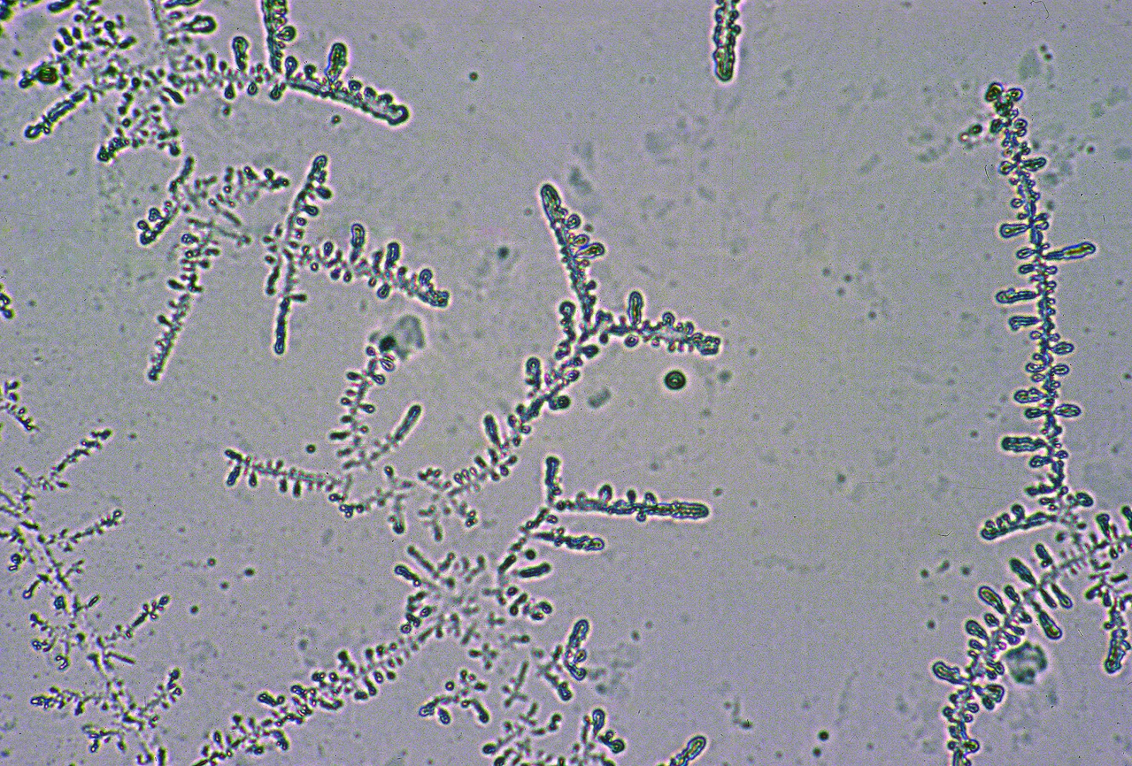
A clear mucous discharge seen at the time of estrus in a cow; an important part of estrous detection in dairy cattle. In some cases, this clear mucous secretion may accumulate in the cranial vagina and be discharged some days after estrus. This mucus contains substantial amounts of sodium chloride (salt). When it is smeared and the salt dries on a glass slide, it generates a so-called "ferning" pattern as seen below.


This mucus is generated by the cervical glands. Therefore, in the case of a persistent hymen, a vaginal mucous discharge is never seen. The mucus accumulates cranial to the hymen where it becomes stained black in color because of metestral hemorrhagic discharges. 

Monday, April 13, 2015

Ovarian activity and cervical mucus in a one month old calf

Keywords: ovary, Graafian, follicle, estrogen, spinnbarkheit

The reproductive tract of a one month old heifer. The tract appeared to be normal; the hymen already perforate.


Image size: 1813 x 1789 px

Although there were no macroscopically visible follicles in the ovaries, numerous small tertiary (Graafian) follicles were present (see inset).  There was also an accumulation of mucus in the cranial vagina showing spinnbarkheit similar to that seen in cows during estrus. This suggested that the follicles were steroidogenically active.

Note:
Postpartum ovarian weight in heifers increases from birth to about 4 months of age but does not increase much more than that until the onset of puberty. During this time, the rest of the reproductive tract also grows, apparently stimulated by the presence of ovarian steroids. Prepuberal (prepubertal) heifers as young as two weeks of age already show regular waves of FSH secretion preceding regular follicle waves; similar to those seen in adults. In fact, both FSH and LH reach substantial concentrations between birth and about two months of age. After that time, negative feedback from ovarian steroids appears to take effect and by three to four months of age the gonadotropins have been suppressed to baseline concentrations. The reason for this early prepuberal rise in gonadotropins is not known.

References:

N.C. Rawlings, N.C. et al. 2003 Antral follicle growth and endocrine changes in prepubertal cattle, sheep and goats. Anim. Reprod. Sci. 78:259–270

Thursday, June 5, 2014

Freemartinism

Keywords: Freemartin, bovine, infertility

The image below shows the reproductive tract from a freemartin heifer. Although there is great variation in the manifestation of Freemartinism, this tract shows some characteristics that are common to many of these animals.


Image size: 1000 x 557px

Note how the development of the Müllerian tubes (precursors of the cranial vagina, cervix, uterus and uterine tubes) was suppressed by anti-müllerian hormone (AMH). AMH was presumably produced by the small gonads seen here where ovaries are usually found. Although it was not verified histologically in this case, these gonads would have contained cells from the male co-twin, including Sertoli-like cells that produce AMH. Of course, it is normal for a male fetus to produce AMH because the Müllerian system must be suppressed if the male is to develop normally. However, in female fetuses where the gene for AMH production is inactive, AMH production by the gonads is disastrous.

As alluded to above, gene coding for AMH production is active in the male co-twin. Therefore gonadal cells derived from the male produce AMH in the co-twin's (Freemartin) gonad. In addition, AMH from the male twin circulates in the female co-twin, amplifying the suppressive effect on the female tract. That has occurred here; the uterus is a mere thread, the cervix is absent and the cranial vagina is also absent. The caudal vagina develops from the ectoderm in a fetus, a system that is not affected by AMH, so it is virtually normal in Freemartins. In this image, a probe has been inserted into the vagina, demonstrating the presence of a normal vestibule i.e. cranially, up to the level of what would have been the hymen. More cranial to that, normal tract is almost absent.

As discussed below, there is a considerable range in the manifestation of Freemartinism;  the tract may be barely affected or may be extremely masculinized. The specimen shown here would probably be considered modal within that range.

Notes: 

In 2005, A.M. Padula  published an excellent review of  Freemartinism (Padula. A.M. 2005. The freemartin syndrome: an update. Anim. Reprod. Sci. 87: 93–109). For additional detail, one should consult that article and others cited within.

At approximately 28 to 30 days of gestation, the allantochorion membranes of most ruminant co-twins fuse. This causes vascular anastomoses to form between the two conceptuses, resulting in freemartinism, a condition that is especially common in cattle. Freemartins occur in other ruminants, cervids and even pigs. However, the condition is comparatively rare in those species (except perhaps for sheep). This author is not aware of any well-documented case in a horse.

Cellular or humoral influences (or a combination of both) circulate between the two fetuses and affect gonadal development. The earlier the fusion, the more masculinized the female tract will be.  In a few cases, the tract is so highly masculinized that it is only slightly ambiguous. The opposite is also true i.e. females can be so mildly masculinized that they are not even recognizable as freemartins, apart from being subfertile.

Interestingly, exchange of cellular material early in gestation also makes freemartins highly tolerant of organ allografts.

Although it is often stated that male co-twins to freemartins are also affected and show deficient spermatogenesis, this is not always so. In fact, males are often completely fertile into adulthood.

On occasion, a male co-twin may die in utero after vascular anastomosis has occurred and although it is unusual (recall that their blood supplies are fused) the female co-twin may survive. In such cases a freemartin may be born as a singlet.

Typically, the Müllerian system in freemartins is severely suppressed; in most cases almost impossible to delineate clearly by transrectal palpation (see the image in this entry). This is because the gonads are a mixture of testicular and ovarian tissue, (freemartins are true hermaphrodites). Sertoli cells in the testicular tissue appear to produce müllerian inhibiting factors as they do in normal males. The gonads are also very small and very difficult or impossible to define on transrectal palpation. The vagina of a freemartin is very short (usually less that 12 cm) and it has a blind ending, the cervix being part of the müllerian system.

Statistically speaking, a female calf born co-twin to a male has approximately a 95% chance of being a freemartin. However one may need further assurance that this is the case. 

In newborn calves, where a transrectal examination is impossible, this author uses the cover sheath of Cassou artificial insemination pipette as a vaginal ‘depth gauge’. The sheath is inserted into the vagina of the suspect freemartin and bent at the level of the vulva lips. A second sheath is inserted into the vagina of a normal calf and bent at the same level. One then compares the length of  sheaths to the point at which they were bent.  The length of the section of interest is far shorter in the suspect freemartin calf than the normal calf. The only chance of an error with this method is in normal heifer calves that have imperforate hymens.

In a farm setting, one may have to wait for a suspect freemartin heifer to reach an age at which she can undergo transrectal palpation safely.

If a diagnosis of freemartinism is still uncertain after considering the history, palpable findings, and external and internal appearance of the heifer, numerous other tests can be performed, mostly of a genetic nature. Sex chromosome chimerism (xx and xy) is present within nucleated blood cells and contrary to older data, in lower numbers in other cells throughout the body as well.

The origin of the term freemartin (also free-martin) is a subject of great uncertainty. As discussed by J.B. Marcum in the Commonwealth Bureau of Animal breeding and Genetics abstracts in 1974 (42: 227) the term may have originated in 1593 when "Martin" was a word used a England and Scotland for a cow or ox. The etymology of the prefix "Free-" is more obscure, possibly arising as a contraction of  'farrow" meaning a barren or non-lactating cow or perhaps the German term "farre" meaning bull, denoting a cow that was like a bull.

Monday, September 30, 2013

Atresia ani. Defecation through vulva


Keywords: atresia ani, calf, bovine


Image size: 5872 x 3640 px

A three-month-old Holstein calf presented for tenesmus and inability to defecate. This animal had been on a milk replacement diet since birth but began to show symptoms when placed on a solid diet. The animal was euthanized. The external appearance of the perineum suggested partial development of the anus and anal sphincter but the absence of any connection with the rectum. Watery feces had passed through a small fistula connecting the dorsal aspect of the vestibule with the rectum. The vestibule was inflamed but the tract cranial to the hymen appeared to be normal.

At three months of age, there was substantial evidence of follicle development on both ovaries.