BOVINE IMAGES

How to use these images


IMPORTANT: To see an enlarged image, click on any image you see in LORI. Then, RIGHT click on the enlarged image to save it at its full size.


Wednesday, July 31, 2013

Estrous mucus discharge in a cow


Keywords: mucus, bovine, estrus, detection, management, dairy, insemination, salt, ferning, crystals


A clear mucous discharge seen at the time of estrus in a cow; an important part of estrous detection in dairy cattle. In some cases, this clear mucous secretion may accumulate in the cranial vagina and be discharged some days after estrus. This mucus contains substantial amounts of sodium chloride (salt). When it is smeared and the salt dries on a glass slide, it generates a so-called "ferning" pattern as seen below.


This mucus is generated by the cervical glands. Therefore, in the case of a persistent hymen, a vaginal mucous discharge is never seen. The mucus accumulates cranial to the hymen where it becomes stained black in color because of metestral hemorrhagic discharges. 

Cervical mucus on the tail of a cow


Keywords: estrus, mucus, management, bovine, dairy, detection


Dried cervical mucus on the tail of a cow. Sometimes this may be the only evidence of a mucus discharge in a cow. In some cattle it is very difficult to detect standing estrus; every possible cue must be taken into account. These include increased activity, decreased milk production, increased vocalizing, mucus discharges from the vulvar lips and after estrus, hemorrhagic metestral discharges.

Bovine abortion of unknown origin

Keywords: bovine, abortion, BVD, IBR, Neospora, diagnosis, retained, placenta, zoonosis


Image size: 1904 x 1308px

Abortion of a bovine fetus at approximately 5 1/2 months of age (estimated on the basis of its crown rump length).  Despite adequate pathological investigation, the etiology was not established. This is frustrating and extremely common; few would contest the statement that "A specific etiology is probably diagnosed in less than 50% of all cases of abortion". Major causes of abortion in cattle include Neospora bovis, Bovine viral diarrhea (BVD), Listeriosis, Infectious bovine rhinotracheitis (IBR), mycotic infections and others, too numerous to mention. Cattle are commonly vaccinated against BVD and IBR to prevent abortion.

Another abortion. Again, the etiology was not determined.


Image size: 1358 x 1229px

This fetus was 218 days old at the time of abortion. After 150 days of pregnancy, retention of the placenta is likely and it becomes progressively more common as the duration of pregnancy increases. In this case, the placenta was firmly attached immediately after abortion (see yellow ring) and was retained for several days. During that time, her body temperature was monitored twice daily. Should the cow have become febrile, antibiotics would have been administered.

Note that the operator is wearing gloves. Although gloves are seldom if ever worn while delivering calves, it is advisable to do so after fetuses are aborted. This is because several abortifacient pathogens are zoonoses. These include B. abortus bovis, Leptospira spp, Chlamydia abortus and  Listeria monocytogenes. Care should be taken to wash thoroughly after handling such cases.

Tuesday, July 30, 2013

Bovine artificial vagina


Keywords: AV, bovine, artificial, vagina, semen, collection


Image size: 1600x 1287px

An artificial vagina used to collect semen from bulls. For diagnostic purposes, semen is usually collected from bulls using electroejaculation but for routine collection of semen, an artificial vagina is used. This artificial vagina was simply made from rigid rubber tubing with a thin, sterile, latex-type rubber liner. Special liners are required because latex itself is often spermicidal, depending on the vulcanizing process used in its manufacture.

Hot water at about 37° C is added to the space between the rigid tube and the liner for warmth, then it is inflated by blowing into the small covered hole seen at left. This is done by lifting the strip of latex covering the hole, blowing into the hole and releasing the strip of latex.

Note the outer jacket surrounding the collection vessel; during cold weather, this is filled with warm water to prevent cold shock of the spermatozoa.

A paraovarian cyst


Keywords: paraovarian, bovine, cow, ovary, cyst, müllerian, wolffian, Kobelts


The structure shown here could either be a wolffian  (Kobelt's or mesonephric) cyst or a remnant of the müllerian system i.e. a paramesonephric cyst. Even in the human literature there is considerable confusion as to the origin of these cysts. Therefore they are often grouped loosely as "paraovarian cysts". Even the histology of the cyst may not reveal its origin, especially in veterinary medicine where these cysts had not been studied as adequately as they have in human medicine.

The cysts have virtually no practical importance because they usually do not impede normal reproductive function. They are occasionally seen on ultrasonography of the ovaries and can be palpated if they are large. 

Paraovarian cysts appear to be more common in horses than in cattle, but in theory, could exist in any species.

The incision across the ovary is incidental; made to demonstrate the presence of the corpus albicans, it's crown just visible at the site of the incision.

Friday, July 26, 2013

Differential interference microscopy (DIC) of bull spermatozoa


Keywords: bovine, BSE, bull , bovine, DIC, interference, microscopy, Nomarski, sperm,


Nomarski Interference Contrast (NIC) or differential interference contrast microscopy (DIC) used here to examine bovine sperm. The advantage of this technique is that no staining is required so live spermatozoa can be examined.

On the left side of the image, two abnormal spermatozoa are present, one with a bent midpiece and the other with a piriform (pear-shaped) head. At lower left and upper right there are two apparently normal spermatozoa. In the middle and bottom of the image, the spermatozoan has a coiled midpiece and tail, reminiscent of the so-called "Dag defect". At lower right there is an enlargement at the end of the midpiece; probably an expanded portion of the mitochondrial sheath. Because this could also be a small midpiece droplet, one could only verify that diagnosis via electron microscopy. The end-pieces of the spermatozoa (the portion of the tail beyond the main piece, at the very end of the tail) are easily seen in this image.
Puberty. Bos indicus

Keywords: Afrikander, Bos indicus, bovine, breeds, puberty, nutrition,


These calves serve as an example for the discussion of puberty, occurring much earlier in Bos taurus var taurus breeds (English and European animal such as Herefords, Angus, Simmental etc) than in Bos taurus var indicus breeds such as Afrikanders, Texas Longhorns, Brahmans etc. Puberty is highly dependent on body weight and therefore, energy intake i.e. if an animal reaches 60% or more of its adult body weight sooner than a herd mate, it will reach puberty sooner.

In females it is often defined as the first time that ovulation and estrus occur together and in males, the first time that a fertile ejaculate is produced.

Normal postpartum bovine uterus.


Keywords: postpartum, suckling, beef, dairy, lochia, involution, uterus, estrus, ovulation


Image size: 1751 x 1131 px

A normal completely-involuted bovine uterus. Note to the typical Y-shaped structure with a short body and two long horns. In the left ovary, a follicle approximately 2 cm in size can be seen developing. In the contralateral (right) ovary, there appeared to be no significant structures. This is typical of a postpartum reproductive tract in a suckled beef cow.. Cows (milk but not suckled) may ovulate as soon as 10 or 14 days after calving, long before the uterus has even involuted.

The corpus luteum and corpus albicans.

Keywords: CL, bovine, cow, palpation, progesterone


Image size: 1104 x 728px

The crown of a corpus luteum (CL) in an ovary of a cow.  The CL is a source of progesterone and several other hormones, including relaxin, müllerian inhibiting hormone, inhibin and even GnRH.

A CL is easily palpable per rectum in most cases and is useful for determining if the cow is having estrous cycles and in some cases, assisting one in staging the estrous cycle. The crown of a corpus luteum is seen in all species except horses where the tunica albuginea does not allow the follicle to ovulate through the surface of the ovary.

After ovulation, blood fills the antrum of the follicle, coagulating and forming a corpus hemorrhagicum. As this blood is phagocytosed, the theca granulosa of the ovarian follicle undergoes both hyperplasia and hypertrophy, folding in towards the center of the antrum.

This image shows the size of a mature CL, the author's thumb depressing the cut surface of that structure in a transected ovary.


Image size 900 x 623px

In the example shown below, that folding is clearly visible.


Image size: 2092 x 1225px

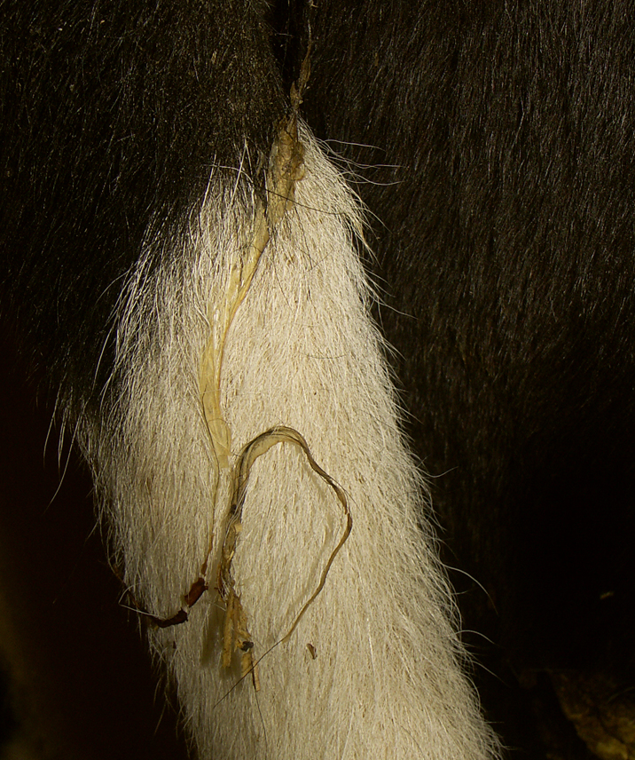
After luteolysis, the corpus luteum undergoes regression and shrinks, forming a corpus albicans. The term "albicans" is derived from the Latin word for white but in domestic animals, this is practically a misnomer. In fact it is only in a few animals (cetaceans for example) where the corpus albicans remains as a large, substantial structure after luteolysis.  In domestic animals it only becomes a small, white body long after luteolysis is complete and in fact, is often invisible to the naked eye by that time.  Indeed, at the end of the luteal phase in domestic animals, when it is commonly referred to as a corpus alicans, the corpus "albicans" (CA) is a dark yellow-to-bright orange structure as shown here.

A CA is seen below, advanced in its luteolysis and degeneration, evidence by rays of fibrous tissue in the structure. Note that this luteal structure has a cavity in the center; a common finding in luteal tissue in cattle. It is not abnormal and is merely a function of luteinization from the theca granulosa growing but never reaching the center of what was previously a follicle. Instead of being referred to as a cystic CL/CA it is often labeled a "luteal cyst".


Image size: 704 x 486px


Metestrus 1: Metestral hemorrhage in bedding.


Keywords:  metestrus, bovine, management, cow, estrous, cycle, detection, hemorrhage


Image size:454 x 326px

This is seen in about 50 to 60% of cows and 75 to 85% of heifers.  It is usually seen in the first 48 hours after ovulation but because the vagina may slope downward at its cranial end (especially in older animals) vaginal mucus and metestral hemorrhage may be retained in the vagina for several days, being discharged several days after estrus. If metestrus is suspected, one should look at the rump of the animal and her dung and bedding where there is often evidence of hemorrhage.

Careful observation is warranted because hemorrhage may be difficult to spot:


Image size: 750 x 526 px

Metestrus 2: Metestral hemorrhage on the tail


Keywords:  metestrus, bovine, management, cow, estrous, cycle, detection, hemorrhage


Image size: 1584 x 2816px

This is seen in about 50 to 60% of cows and 75 to 85% of heifers.  It is usually seen in the first 48 hours after ovulation but because the vagina may slope downward at its cranial end (especially in older animals) vaginal mucus and metestral hemorrhage may be retained in the vagina for several days, being discharged several days after estrus. If metestrus is suspected, one should look at the rump of the animal and her dung and bedding where there is often evidence of hemorrhage.

Metestrus 3: Metestral hemorrhage on the tail 

Typical metestral hemorrhage; seen on the tail of a cow.


Image size: 1584 x 2816px

This is seen in about 50 to 60% of cows and 75 to 85% of heifers.  It is usually seen in the first 48 hours after ovulation but because the vagina may slope downward at its cranial end (especially in older animals) vaginal mucus and metestral hemorrhage may be retained in the vagina for several days, being discharged several days after estrus. If metestrus is suspected, one should look at the rump of the animal and her dung and bedding where there is often evidence of hemorrhage.

Metestrus 4. Hemorrhage seen in bedding 

Typical metestral hemorrhage; seen on the bedding of a cow.


This is seen in about 50 to 60% of cows and 75 to 85% of heifers.  It is usually seen in the first 48 hours after ovulation but because the vagina may slope downward at its cranial end (especially in older animals) vaginal mucus and metestral hemorrhage may be retained in the vagina for several days, being discharged several days after estrus. If metestrus is suspected, one should look at the rump of the animal and her dung and bedding where there is often evidence of hemorrhage.

Metestrus 5. Hemorrhage seen in bedding

Keywords:  metestrus, bovine, management, cow, estrous, cycle, detection, hemorrhage


Image size:1164 x 774px

This is seen in about 50 to 60% of cows and 75 to 85% of heifers.  It is usually seen in the first 48 hours after ovulation but because the vagina may slope downward at its cranial end (especially in older animals) vaginal mucus and metestral hemorrhage may be retained in the vagina for several days, being discharged several days after estrus. If metestrus is suspected, one should look at the rump of the animal and her dung and bedding where there is often evidence of hemorrhage.

Metestrus 6. Hemorrhage seen in the uterus

.


This image shows a remarkable example of hemorrhage into the lumen after ovulation; an effect on the permeability of endometrial capillaries due to estrogen withdrawal. A similar phenomenon is seen in humans shortly after ovulation but is seldom noteworthy. Note the CH on the left ovary from a recent ovulation and the CA from the previous cycle on the right.

Metestrus 7. Hemorrhage on the rump of a cow.

Keywords:  metestrus, bovine, management, cow, estrous, cycle, detection, hemorrhage


Image size:1584 x 2816px

Typical metestral hemorrhage; seen on the rump of a cow that was recently in heat. This is seen in about 50 to 60% of cows and 75 to 85% of heifers.  Metestral hemorrhage is usually seen in the first 48 hours after ovulation but because the vagina may slope downward at its cranial end (especially in older animals) vaginal mucus and metestral hemorrhage may be retained in the vagina for several days, being discharged several days after estrus. If metestrus is suspected, one should look at the rump of the animal and her dung and bedding where there is often evidence of hemorrhage.

Cattle are the only domestic animals that show hemorrhage as a result of estrogen withdrawal.

Kamar heatmount detector

Keywords:  Kamar, bovine, management, cow, estrous, cycle, detection


The Kamar device is applied to the rump of a cow with adhesive. When the cow is in standing estrus and allows another cow to mount her, a chamber containing dye is crushed, turning the device red throughout its length. Although valuable, the device is prone to false positives in crowded barns when cows are moving in or out of the building; in such cases mounting may be less specific than otherwise.

Cystic follicle in a cow


Keywords:  anestrus, COD, cyst, cystic, follicle, bovine, management, cow, estrous, cycle


Reproductive tract from a cow with cystic ovarian disease (COD); a syndrome associated with high milk production and negative energy balance in high producing dairy cows. COD is usually associated with persistence of large, un-ovulated follicles in the absence of standing estrus, probably because of luteinization (with progesterone production) of the cystic structures due to low pulsatile releases of LH. Nevertheless, this illustration shows that ovulation occasionally occurs in the presence of a cystic follicle. Note the corpus luteum in the left ovary, in the same ovary as the cyst.

In these cases, the cyst may then regress and may even be replaced by other cysts until ovulation becomes predictable and cyclicty returns to normal.

This illustration also suggests that luteinization of cystic follicles may be so limited that they not produce enough progesterone to suppress LH and block ovulation.

The terminology of COD can be confusing, with cystic follicle occasionally being referred to as luteal cysts. In the author's opinion this term should not be used because it can be confused with a structure called a cystic corpus luteum; a non-pathological entity. Instead, when a cyst becomes luteinized it should be called a luteinized cyst rather than a luteal cyst.

Hydrops in a cow


Keywords:  hydrops, bovine, cow, IVF, cloning, allantois, amnion


A cow with both hydrops amnion and allantois, showing her enlarged abdomen. It is sometimes stated that hydrops amnion develops slowly and in the presence of an abnormal calf. In the case of hydrops allantois, some maintain that the hydroptic condition develops more rapidly and the calf is usually normal. However there is often overlap between the two conditions with hydrops developing in both fetal placental compartments. Hydrops has become more common in cattle with the advent of in vitro fertilization and cloning.

In Japanese Black cattle it has been shown that hydrops allantois can be associated with a recessive gene coding for hydronephrosis and polyuria; this results is hydrops allantois.

Selected reference:

Sasaki S. et al. 2016. A missense mutation in solute carrier family 12, member 1 (SLC12A1) causes hydrallantois in Japanese Black cattle BMC Genomics 17:724 739

Thursday, July 25, 2013

Species placentation


Keywords: placentation, placenta, species, comparative, anatomy


Image size: 1000 x 1375px

An amalgam of images showing the gross anatomy of the placentas of various domestic species.  All of these occur in the domestic animals but many other forms occur in other animals. For example, primates and humans have a discoid type of placentation; more intimate than any in domestic animals.

In image A, a multiplex type placenta is illustrated. In this case, it is the placenta of a ewe; similar to that of a cow (seen in image B) with subtle differences.

Image C from a neonatal calf, shows that the sites of the caruncles are established very early in the life.

Image D shows the zonary placenta of a dog; similar to that of a cat, shown in image E.

The placentas in F, G and H are all diffuse in character: Image F shows the placenta of a piglet, G the placenta of a foal and H, the placenta of a Llama, a new world camelid. Interestingly (although they are similar to ruminants) all of the camelids have diffuse placentas, not the multiplex type of placentas that one finds in domestic ruminants.

A bovine granulosa cell tumor. 


Keywords: bovine, GCT, granulosa, tumor, neoplasia, cow


Image size: 5184 x 3456px


Image size: 5184 x 3456px

A tumor in the ovary of a cull cow. This was a slaughter plant specimen with no other history. Histopathology confirmed that this was a granulosa cell tumor (GCT). That histopathology is shown in another LORI entry

Sex cord tumors of the granulosa-cell type are the most common sex cord-stromal tumors in cattle. They occur in all age groups, even in prenatal and very young calves, and in all breeds.

Steroid production has been reported but is not well characterized. Some studies show that they can produce estradiol 17 beta, progestogens and testosterone. Mammary enlargement in non-gravid affected heifers also suggest that estrogens are produced by some of these tumors.   Those findings are not surprising in terms of the de-differentiated nature of cells that normally produce steroids.

As is the case in other domestic animals  (all farm and companion animals) where granulosa cell tumors have been described, systemic metastasis is not typical but has been described in most species.

Some granulosa cell tumors in cattle can be cystic or polycystic with granulosa cells lining the cysts while others, such as this one, are solid with solid types probably in the majority. As the tumor outgrows its blood supply, areas of necrosis and hemorrhage may occur. Histologically, the neoplastic cells may be in trabecular, diffuse patterns or follicular.

Neoplastic cell types in these tumors have been described as granulosa- like or Sertoli cell- like with cells of either type predominating in separate tumors.

In this case, the tumor weighed approximately 10 kg, somewhat smaller than the largest described at 17.1 kg.

Although granulosa cell tumors such as this one may resemble Theca Cell tumors (also solid and yellow to orange in color)  Theca cell tumors consist of masses of spindle- like cells almost resembling fibromas. They are not nearly as common as granulosa cell tumors.