BOVINE IMAGES

How to use these images


IMPORTANT: To see an enlarged image, click on any image you see in LORI. Then, RIGHT click on the enlarged image to save it at its full size.


Showing posts sorted by date for query pregnancy. Sort by relevance Show all posts

Thursday, November 7, 2019

Technique for artificial insemination; bovine.

Keywords:  bovine, vagina, insemination, A.I., AI, technique.

Routine artificial insemination (AI) in cattle is more complex than routine AI in mares.  The common method of AI in mares usually involves non-frozen semen. A gloved hand is simply inserted into the vagina then a finger into the cervix, enabling guidance of an insemination pipette into the uterus. In cattle, semen is almost invariably frozen and must be thawed carefully then inseminated by guiding an insemination rod through the cervix using transrectal manipulation. The bovine vagina, especially in heifers, will not permit the easy entry of a gloved hand and certainly, a finger cannot be passed through the cervix of a normal non-pregnant cow or heifer.

The intent of this entry is to illustrate the mechanics of handling an insemination rod and the act of insemination itself. The author is indebted to Select Sires US & Canada for allowing these images to be used in LORI. Select Sires holds the copyright to all images in the entry and should be contacted regarding their use outside of this image library.

After removing most of the feces from the rectum, the gloved hand is used to grasp the cervix transrectally. Then, using paper towel, the vulva lips are cleaned briefly, and the cow's tail is deflected to one side using the arm in her rectum. The vulva lips are parted (if a helper is not available, one of  the techniques described in the text box below can be used) and the AI gun is inserted into the vestibule at an upward angle. Obviously, care must be taken not to contaminate the vagina. See figure 1. It is important to grasp the cervix and push it cranially to eliminate vaginal folds that often impede passage of the AI gun.


Figure 1. An AI gun is inserted to the level of the external cervical os. In the author's opinion, the hand shown here should advance cranially to hold the entire cervical body. Holding the entire cervix in one's hand allows it to be manipulated effectively. At this point, the author also advances the AI gun so that it can be felt against the fifth digit (small finger) in the ventral part of the cervical fornix. With the entire cervix is within one's grasp, the external cervical os is lifted and fed over the tip of the AI gun. Image size:1000 x 680

Variations: One can hold the base of the loaded insemination between one's teeth; a valuable "third hand". The paper towel that has been wrapped around the AI gun to prevent semen cold shock, is removed. Some technicians fold this paper towel, kink it and place it in the ventral vulva commisure. This opens the vulva lips and serves as a surface upon which to slide the AI gun upward and into the vagina. In any case, the vulva lips should be wiped as clean as possible; generally not repeating this action without more paper towel, as that leads to more contamination than otherwise. The author uses a different approach, pushing down slightly on the cleaned perineal body using the elbow of the arm in the rectum. This causes the vulva lips to gape while the AI gun is inserted into the vagina.
  

Figure 2. An approach where the cranial vagina is narrowed over the external cervical os to facilitate entry of the AI gun into the external cervical os. As stated above, the author uses a different technique to locate an entry point for the AI gun. Although the external cervical os can also be located using an endoscope designed for this purpose, this does not necessarily facilitate passage of the AI gun through the cervix, often the most challenging part of AI in cattle. Image size:1000 x 680

Using either technique, after the AI gun has entered the cervical canal, the rings of the cervix are manipulated over the tip of the AI gun.  See figure 3. Note the emphasis on manipulating the cervix over the gun and not placing primary emphasis on moving the gun cranially, attempting to bypass the cervical "rings" (they are not true rings but a series of crescents). The AI gun should not be forced cranially in hopes of displacing the annular rings .

Figure 3: Manipulating the annular rings (folds) of the cervix over the AI gun as it is moved gently towards the uterine body. Image size: 800 x 554

Although it is not illustrated in this entry, it is essential to place a finger over the cranial aspect of the cervix to determine that the AI gun has emerged from the internal cervical os and lies within the uterine body. The plunger of the AI gun is then depressed, expelling semen into the uterus (see figure 4). One should ensure that the AI gun is not pulled caudally into the cervix while the plunger is depressed.


Figure 4. Depositing semen within the uterine body. Image size:1000 x 680

Clitoral stimulation in both cows and heifers immediately after insemination has been shown to increase pregnancy rates marginally in some studies but this effect (especially in heifers) is not consistent. 

Some studies show advantages to intra-cornual (in the uterine horns) insemination, others not. The situation remains unclear. For routine AI therefore, it is reasonable to suggest that semen should be deposited within the uterine body (the short section of the uterus that extends only 2 to 3 cm cranial to  the internal cervical os). On the other hand, there appear to be no detrimental effects when intra-cornual insemination is used. Therefore intra-cornual should be considered when sexed semen is used or when super-ovulation is practiced.  Passing an AI gun up the uterine horn requires different techniques to those shown here, akin to those used for embryo collection. 

Selected references:

Carvalho P.D. et al. 2013 Effects of deep-horn AI on fertilization and embryo production in superovulated cows and heifers. Theriogenology. 80:1074–1081

Ciro, M. et al . Comparison between deep intracornual artificial insemination (dIAI) and conventional artificial insemination (AI) using low concentration of spermatozoa in beef cattle. Braz. arch. biol. technol.[Internet]. 2012 June.cited 2019. 55: 371-374.

Lunstra, D.D. et al. 1983. Clitoral stimulation and the effect of age, breed, technician, and postpartum interval on pregnancy rate to artificial insemination in beef cattle. Theriogenology. 19:555-563

Momont, H. et al. 1989 Does intrauterine site of insemination in cattle really matter? Theriogenology 32:19-26

Segura, C.V.M. and Rodriguez, R.O.L. 1994. Effect of clitoral stimulation after artificial insemination on conception in Zebu-crossbred heifers in the tropics. Theriogenology 42:781-787

Saturday, November 24, 2018

Uterine torsion

Key words: uterus, torsion, bovine, accident, pregnancy


Uterine torsion is not uncommon in cows. It is seldom encountered in heifers and is also infrequent in Bos taurus var indicus cattle. The reasons for those findings are not clear but have been related to large, deep and expansive abdomens in affected breeds and parity. Uterine torsion is also more common in unfit dairy cows kept in barns than those at pasture; perhaps a consequence of poor abdominal muscle tone and weak limbs allowing sudden jerky movements.  Not surprisingly, uterine torsion has also been related to poor uterine tone and hypocalcemia.

One only has to appreciate how the hind quarters of a cow are elevated above its thorax when it rises or lies down to imagine how the stability its pendulous uterus becomes precarious in that posture.  The inherently poor stability the bovine uterus adds to this predisposition. Consider the attachment and flaccidity of the mesometrium in cattle. This allows one to easily retract and manipulate a non-pregnant bovine uterus during transrectal palpation. During pregnancy the mesometrium offers even less support as the pregnant uterus expands cranially, beyond the cranial margin of the mesometrium.

In twin pregnancies, the uterus is more evenly loaded and is a result, more stable. Not surprisingly, uterine torsion is comparatively rare in cows with twin pregnancies.

Uterine torsion occurs in all ruminants and even multiparous animals but is most common in cows and horses, especially cows. In horses, mesometrial support is far more substantial than it is in cattle. One only has to try to retract an equine uterus to appreciate that reality. Even during pregnancy, the equine uterus is more stable than a bovine uterus. Also, mares change from the lying to standing posture by raising their forequarters first, so the equine uterus seldom finds itself in a pendulous state. The cause of uterine torsion is different between the two species; in cattle, as explained, it is due to uterine instability during early parturition. In mares it appears to be related to mares rolling in pasture during mid to late gestation; perhaps as they shed their winter coats.

In this entry, all descriptions of the direction of torsion are written as if one is looking at the cow, caudal-to-cranial. In that regard, anti-clockwise torsion is more common than vice versa. The reason for this is not obvious and the author's speculation will not add to the value of this entry. In the case discussed here, torsion was in a clockwise direction.

Figure 1: The mechanism of uterine torsion in cattle. In this illustration the uterus has twisted clockwise, pulling the left mesometrial ligament (LM) over to the right, cranial to the right mesometrial ligament (RM). The arrow indicates the direction of torsion. Size available: 900 x 551px

This illustration is found elsewhere in various manifestations on the Internet. However, the original image was drawn by Dr Ronald Trengrove circa 1971. Shortly before his death in 2014, Dr Trengrove gave the author permission to use and edit his veterinary drawings. 
font-weight: normal>

In rare cases, uterine torsion can occur during mid gestation but it is usually a condition associated with calving. Typically, a cow affected with uterine torsion will show cessation of calving but continuing signs of discomfort. The farmer notes the typical mucous discharge from the vulva (see figure 2) the presence of colostrum,  and obvious signs of impending  parturition. Then there is no further progress. After several hours of inactivity but persisting discomfort, a veterinarian is called.

Figure 2: A pluriparous cow with uterine torsion, several hours after the onset of calving. Note the copious clear vulva discharge and distended udder. In this case, there was no obvious distortion of the vulva lips; an occasional finding in these cases. Size available: 1149 x 1159px.

If the fetus or its membranes have entered the vagina, there may be a degree of straining.  On transrectal examination, a tight mesometrial ligament can be felt stretching across the caudal abdominal cavity. Torsion of the uterus may be palpable per rectum as a corkscrew to the left or right-hand side. On vaginal examination, torsion is usually detected, but in a few cases, the site of torsion is cranial to the cervix and vaginal torsion may be absent. If the cervix has dilated, fetal limbs may be in the vagina. Often however, uterine torsion occurs before the cervix has dilated completely. If torsion is not relieved, fetal death, putrefaction and toxemia will result.
.
Figure 3: An operator (Dr J. Spears) determining  the severity of torsion and patency of the cervix.  Perhaps obviously, the cervix was indeed patent in this case; open enough to allow the operator to place a lubrication tube and calving chains. As a result rolling (described later in this entry) was not required. Size available: 1600 x 1200px

Figure 4: This image shows two obstetrical chains and a Cornell detorsion rod about to be used to rotate the calf in a clockwise direction. Size available: 1175 x 1172px

Figure 5: In this case the operator has elected to use a Cornell detorsion rod to rotate the fetus. The two red arrows in the inset image show how the calves legs are placed through loops of the calving chains prior to detorsion. The inset shows how a single chain can be use with the detorsion rod. In this case however, the operator chose to use two chains with the detorsion rod. Size available: 2203 x 1556px

A plastic device named "Gyn-Stick", similar to the Cornell detorsion rod, is available from Jorgensen labs.  It is made from rigid plastic and uses calving ropes in place of chains. Its use is shown in this commercial video.

The author was introduced to  the Cammerer's detorsion fork as an undergraduate in 1972. It is shown in figure 5, a diagram by the author. The commercial product can be seen on this website, still commercially available. In the author's opinion it is preferable to Cornell-style instruments because large cuffs spread the torsion load on the limbs and the fork enlarges the radius of rotation compared to other detorsion instruments.

Figure 5: A Cammerer's* detorsion fork' often referred as a torsion fork. Size available: 624 x 1132px

Figure 6: After successful detorsion, the cervix of the cow was dilated manually over a period of 15 to 20 minutes prior to extraction and the calf. The obstetrics chains were moved distally from their original positions on the left and right proximal radius and ulna; they were placed on conventional traction sites, above each fetlock joint. As is usually the case, the chains were also thrown into a half hitch proximal to each pastern joint.  Size available: 1409 x 1119px

Figure 7: Torsion was then used to rotate the calf by 110 to120 degrees into a dorso-left-ilial position to prevent hiplock; a normal part of mutation and traction in any assisted calving. Size available: 1600 x 1200px

Figure 8: The calf was dead in this case probably because torsion was protracted and severe. In one large study (Klaus-Halla, D. et al. 2018) 35 percent of calves were delivered live if the case was treated within 12 hours after the onset of calving and approximately 90% if treatment began within 6 hours. Size available: 942 x 1461px

In some cases the cervix has not yet dilated at the time of presentation or the uterus has twisted to such an extent as to preclude entry by the operator. In those cases, the cow must be rolled to relieve torsion. After torsion is relieved, it is possible to determine if the cervix and body of the uterus can be accessed to deliver the calf. Fortunately, in the majority of cases, the cervix will have dilated enough to permit per vagina mutation.

In the opinion of some, if cervical dilation is insufficient to allow per vagina delivery after rolling, one should wait for at least three hours for this to occur. However, the cervix may never dilate sufficiently in some cases and even if the cervix does dilate, a second visit to the farm will be required at extra cost; only to deliver a dead calf. In the author's opinion therefore, it is preferable to perform a Cesarean section immediately after rolling in those cases.

Even if the cervix is closed and it appears impossible to deliver the calf per vagina, the cow should be rolled to correct torsion. This will facilitate Cesarean section if it is required. In the event that a Cesarean is attempted before torsion is corrected, the uterus, once freed of the weight of the calf, may contract and rotate away from the surgeon. This makes the uterine wall difficult to suture.

A highly experienced colleague responded to this statement by saying that his standard approach to torsion is to perform a cesarean section immediately in all cases of torsion.

The technique of "rolling" is shown in figure 9.

Figure 9: Correction of uterine torsion by rolling (Schaffers’* method). The uterus has twisted clockwise in this case. This is shown by the inner, circular red arrow “a”. An attempt is made to roll the cow in the same direction (large red arrow) as the uterus has twisted so that the cow twists around her own uterus. A plank is often used to facilitate this process with pressure applied to the cow's abdomen, just cranial to her udder. During rolling, pressure on the uterus helps to prevent rotation of the twisted uterine horn. Although the affected uterine does not rotate during rolling, it could be said that it rotates anticlockwise relative to the cow. In this case that would be in the direction shown by the circular green arrow “b”.  If  rolling successful, fetal fluids usually escape from the cow's vulva lips immediately after torsion is relieved. An attempt should then be made to mutate and extract the calf. Size available: 1200 x 894px

*The author has been unable to determine the exact origin of the eponym Cammerer  Any assistance in that regard will be appreciated. Schaffer published on his plank modification for uterine torsion in 1946 (see references). 

Selected references:

Frazer, G.S. 1996. Bovine uterine torsion: 164 hospital referral cases. Theriogenology. 46:739-758

Klaus-Halla, D. et al. 2018 In German. Translated: [Uterine torsion in cattle: Treatment, risk of injury for the cow and prognosis for the calf] Tierarztliche Praxis. 46:143-148 

Lyons N et al. 2013. Clinical forum: Bovine uterine torsion. Livestock. 18: 18-24

Pascale, A. et al 2008. A study of 55 field cases of uterine torsion in dairy cattle. Can Vet J. 49:366–372

Roberts. S.J. Veterinary obstetrics and genital diseases (Theriogenology). Published by the author. Uterine torsion. pp 230-233

Roelofsen, J.M.P. 2018 Uterine Torsion in relation to blood calcium concentration in dairy cattle. Masters Thesis University of Utrecht.

Schaffer W. 1946. Schweizer Arch. Tierheilk 88: 44.


Thursday, November 30, 2017

Uterine endometrial fissures

Key words: bovine, endometrium, fissures, uterus, postpartum, pyometra

The opened uterus shown below (image 1) was obtained from a seven-year-old Holstein cow in poor body condition. She was euthanized and submitted for postmortem examination because of infertility and continual loss of weight. Her history of postpartum estrous cycles was unknown.


Image 1, size available: 2005 x 1350 px

Apart from mild subacute suppurative broncopneumonia, the most noticeable feature in this cow was her moderately enlarged uterus, especially the right uterine horn. The uterus was turgid and distended with a large volume (perhaps 2 liters) of cloudy, tan colored, watery pus. The cervix was closed, not allowing any passage of the purulent material into the vagina. 

It is possible that pyometra had been supported by a corpus luteum (CL) formed from a postpartum ovulation (the common situation in pyometra) because a corpus albicans (CA) was present in her left ovary. However, the CA formed from the regression the CL of pregnancy can persist for extended periods after calving so that possibility is questionable. Therefore and most likely, one is left with the remarkable situation where the cervix was closed as is the case in common pyometra, yet there was an absence of luteal tissue to support that condition. More remarkable however, were the multifocal deep fissure-like ulcerations on the endometrium (image 2). These are shown, partially covered by purulent exudate in the right half of the image above. The largest of these measured 2 cm at its widest point. 


Image 2, size available: 3005 x 1773px

As indicated by the yellow arrows in the image above, the fissures were visible from the peritoneal surface as well. The serosal surface of the uterus was covered with a fine layer of yellowish fibrin, indicating an extension to local peritonitis.

Histopathology showed multiple small abscesses within these fissures. Neutrophils were the predominant cell type, accompanied by a low population of mononuclear cells. Culture of the uterine exudate produced no microbial growth; not particularly rare in a purulent environment.

Fissures such as these are not characteristic of common cases of pyometra due to Trueperella pyogenes (formally assigned successively to the the genera Corynebacterium, Actinomyces, then Arcanobacter). Despite an interest in reproductive pathology spanning many years, this author is unaware of similar cases and solicits the input of colleagues in this regard. Certainly, partial circumferential-splitting of the endometrium is a feature usually not seen in pyometra, even in uteruses distended with large volumes of pus.

Friday, April 21, 2017

Late gestation pregnancy and placentation

Keywords: bovine, calving, partus, parturition, placenta, caruncles, placentomes, cotyledons, cervix

These images show some of the characteristics of a pregnant uterus removed from a Simmental cow that died suddenly during the early stages of calving. The cause of death was not determined.

As shown below, the calf was located in the right uterine horn. In that regard, it is useful to remind readers that the uterine horn occupied by the fetus is of little significance during cesarean sections. This is because the so called "non-pregnant" horn contains such a small volume of fetal fluid that it is deflected towards the midline by the horn that contains the calf. Therefore (in a normal pregnancy) albeit left or right, the "pregnant" horn is readily accessible from a normal left flank approach.


Image size: 2888 x 1585 px 

In the image above, arrow A indicates that if one were to raise the right uterine horn and inspect the mesometrium (arrow B) the uterine artery would be visible. From about 4 months of gestation, the volume of blood flow in this artery excedes the ability of its thin wall to constrain blood flow in a linear fashion. In bovine pregnancies, this results in turbulent blood flow, reminiscent of air flowing through a "wind sock". In this video, the author illustrates turbulent air flow that is akin to blood flow vibration felt during palpation. This phenomenon is known as fremitus (< Latin. "To murmur"). Fremitus appears later in the vessel supplying the "non-pregnant" horn than that supplying the "pregnant" horn. In both vessels however, it ceases shortly after calving. Therefore it can still be present after calving and in the strictest sense, cannot be regarded as an infallible indicator of pregnancy.

Interestingly, fremitus is not detectable in pregnant mares.

As shown below, the corpus luteum (CL) of pregnancy (ringed in green) was still present in this specimen, shortly before calving would have occurred. This is normal.


Image size: 1081 x 710 px

Although the CL of pregnancy is still present at term, most cows remain pregnant even if total ovariectomies are performed within the last 30 days of gestation. Indeed, some cows becomes independent of CL progesterone secretion as early as 6 months of gestation. After ovariectomy however, pregnancies do not end normally. Although pregnancies are maintained to term, cervical dilation, dystocia and retained fetal membranes are common. This is because the corpus luteum is a also a source of relaxin, prostaglandins E1 & E2 and even oxytocin. It is therefore, essential for normal parturition.

Soon (12 to 16 hours) after substantial uterine contractions begin, the cervix relaxes rapidly. This relaxation is largely under the effect of PGE2 but as mentioned earlier, other hormones are also involved.

The initial phase of cervical dilation is largely passive i.e. independent of the dividing force of the fetal head and forelimbs. However, the effect of the fetus can not be excluded completely because the uterus is pushing it towards the cervix, even during the early stages of cervical dilation. Within 8 to 10 hours in cows and somewhat longer in heifers, the cervical canal dilates to a diameter of 12 to 20 centimetres i.e. big enough to accommodate the head of the foetus.


Image size: 1000 x 772 px

In the specimen discussed here, passive dilation was incomplete yet almost sufficient to accommodate a human hand (about 12 cm wide, including the thumb).


Image size: 3111 x 1729 px

Normally, in pre-term pregnant cows, even in autolysed specimens, this would be impossible. Therefore had this cow lived, the second (expulsive) stage of calving would have been imminent.

The image below confirms the fact that the fetus had not yet entered the birth canal i.e. active dilation of the cervix and second stage parturition had not yet begun.


Image size: 2138 x 1056 px

The following image shows the fetal membranes and the calf, removed from the uterus.  The normal number of placentomes in bovine pregnancies varies between 75 and 120, the largest measuring 12 to 14 cm in length. Also, as reviewed elsewhere, amnionic fluid volume varies between 2 and 8 liters and allantoic fluid, between 4 and 15 liters. This pregnancy appeared to be normal in those respects.


Image size: 3456 x 2130 px

Placentation in ruminants consists of numerous complexes. Hence the term "multiplex placentation" as apposed to "diffuse, discoidal, zonary" etc.  The two parts of each complex are of course, a caruncle on the maternal side and a cotyledon on the fetal side. Together, they form a placentome. A placentome is shown here:


Image size: 1200 x 809 px

As illustrated elsewhere in LORI, the site and number of caruncles in a cow has already been established when the cow herself was a fetus!

Note how the fetal villi are sliding out of the maternal crypts, a process that is impossible if  placental maturation (flattening of maternal epithelium and loosening of fetal-maternal contact) has not occurred during the last few days of gestation. Incomplete placental maturation is a cause of retained placenta. Placental maturation is impaired if the fetus is born prematurely or the diet of the cow is deficient in selenium and vitamin A, nutrients that are essential for epithelial function. This explains in part why retained placenta is common when selenium and vitamin A are deficient in the diet of pregnant cattle.

Saturday, December 31, 2016

Fetal ascites & anasarca (hydrops fetalis)

Keywords: bovine, anasarca, ascites, dystocia, heritable, neoplasia, hypoplasia, IVF, clone, cloning

Ascites: < Greek askos; "a bag of wine"
Anasarca: < ana : "throughout" & sarca: "new flesh"
Hydrops fetalis: Abnormal accumulation of fluid in two or more fetal compartments, including ascites, pleural effusion, pericardial effusion, and skin edema.

A complex subject with varying appearances and many different etiologies. Hydrops fetalis (HF) may be due to any factor/s that cause passive venous congestion i.e. intra-peritoneal or intra-thoracic neoplasia (leiomyomas, teratomas hepatoblastomas), liver cirrhosis or other liver anomalies, cardiac anomalies, pulmonic valve stenosis or dysplasia of the lungs themselves. Some of these anomalies are heritable in several breeds of cattle. As is so often the situation with abnormal calves, the calf shown below (photographed 41 years ago!) was discarded without a thorough post mortem examination. Therefore the cause of ascites in this case was unknown.

Because of its enlarged abdomen and resulting dystocia, the calf (weighing 71 kg) was delivered by cesarean. A calf with ascites can often be delivered per vagina after incising its abdomen using a finger knife and allowing the fluid to drain into the cow's uterus. The yellow staining  of the hair coat here was due to meconium discharge from the anus; a common situation during fetal distress precipitated by hypoxia.


Image size: 1330 x 891 px. 

In the fetus below (delivered by cesarean and also discarded without a thorough postmortem examination) there was ascites as well as marked anasarca. The calf showed a multitude of musculo-skeletal deformities too.


Image size: 1500 x 818 px

In fetuses produced by assisted reproductive technology (ART) i.e. cloning through nuclear transfer (NT) and in vitro fertilization (IVF), pregnancy failure is common and many abnormalities have been described in calves, lambs and human babies produced through ART. The most striking of these is the so called "Large offspring syndrome" where newborn babies, calves and lambs can be several times their normal weight at birth. Cardiac, kidney, hepatic and pulmonary abnormalities have been described in these cases. In addition, angiogenesis itself can be abnormal, leading to increased permeability of blood vessels in the fetus and fetal membranes.

In the case shown below, the calf was a product of IVF and abortion occurred at 7 months of gestation. There was severe ascites and anasarca (HF) as well as hydrops amnion and allantois. The case is discussed in more detail in another LORI entry, featuring a video. In that video, the author suggests that genetic alterations may occur in these cases as a result of culture conditions. However, current information shows that the abnormalities manifested in cases of IVF and NT are largely epigenetic in nature.

The calf weight 56Kg; well in excess of the 8 to 18Kg range for a fetus of this gestational age. Meconium staining of the hair coat is obvious in this case as well.


Image size: 1333 x 1076 px

Pitting edema (below) was present over the entire surface of the calf and its fetal membranes.


Image size: 1591 x 988 px

Subcutaneous edema is visible here, in the upper left inset. At lower right, a stream of ascitic fluid  is being released from the abdomen. Readers are encouraged to view the teaching video mentioned above to appreciate the nuances of this case.


 Image size: 1677 x 1190 px

In conclusion, ascites, anasarca, placental edema, hydrops allantois and hydrops amnion are common manifestations of  various conditions that may or may nor be related to one another.

Selected references:

Alberto, M.L.V. et al. 2012. Development of bovine embryos derived from reproductive techniques, Reproduction, Fertility and Development. 25: 907-917

Baraya, Y.S. et al. 2015. Dystocia associated with foetal anasarca (hydrops fetalis) in a Nigerian breed of sheep; Case report. Proceedings: Sixth Pan Commonwealth Vet.Conf.of the CVA and 27th Vet. Assn. Malasia Conf. 519-522

Buchanan, J.W. 2001. Pathogenesis of single right coronary artery and pulmonic stenosis in English Bulldogs. J. Vet. Intern. Med. 15: 101-104

Chen, Z et al. 2013. Large offspring syndrome. A bovine model for the human loss-of-imprinting overgrowth syndrome Beckwith-Wiedemann. Epigenetics 8:591–601

De Vries, C. et al 2012. Congenital ascites due to hepatoblastoma with extensive peritoneal implantation metastases in a premature equine fetus. J. Comp. Path. 2012, 148: 69-69

Drost, M. 2007. Complications during gestation in the cow. Theriogenology. 68: 487-491

Edwards, J.L. et al. 2003. Cloning adult farm animals: A review of the possibilities and problems associated with somatic cell nuclear transfer. A. J. Reprod. Immunology. 50: 113-123

Golladay, E.S. and Mollit, D.L. 1984. Surgically correctable fetal hydrops. J.Pediatric Surgery.19: 59-62

Pinborg, A. et al. 2014. Large baby syndrome in singletons born after frozen embryo transfer (FET): is it due to maternal factors or the cryotechnique? Hum. Reprod. 9: 618-27

Pushp, M.K. et al. 2016. Dystocia in a non-descript cow due to ascetic fetus- a case report. J. Livestock Sci 7: 62-64

Rheuban, K.S. et al. 1991. Intrapericardial teratoma causing nonimmune hydrops fetalis and pericardial tamponade: A case report. Pediatric Cardiology. 12: 54–56

Roberts, S.J. 1986. Veterinary obstetrics and genital diseases. Diagnosis and treatment of various types of dystocia. pp 333-335. Published by the author, S.J. Roberts.

Sasaki S. et al. 2016. A missense mutation in solute carrier family 12, member 1 (SLC12A1) causes hydrallantois in Japanese Black cattle BMC Genomics 17:724 739

Svara, T. et al. 2016. Pulmonary hypoplasia and anasarca syndrome in Cika cattle. Acta Vet. Scand. 58: 36-40

Testoni, S.et al. 2009. Congenital facial deformities, ascites and hepatic fibrosis in Romagnola calves. Vet.Rec. 164: 693-694

Walker, S.K. et al. 1996. The production of unusually large offspring following embryo manipulation; concepts and challenges. Theriogenology 45: 11l-120 

Whitlock, B.K. et al. 2008. Heritable bovine fetal abnormalities. Theriogenology. 70: 535-549

Windsor, P.A. et al. 2006. Hydrops fetalis associated with pulmonary hypoplasia in Dexter calves.
Australian Vet. J. 84: 278-281

Young, L.E. et al. 1998. Large offspring syndrome in cattle and sheep. Reviews of reproduction. 3: 155-163

Saturday, December 3, 2016


Guidelines for aging bovine pregnancies.

Keywords: hand, measurement, abortion, pregnancy, diagnosis, bovine, placentomes, crown-rump

It is not the author's intention to foist a personal bias onto colleagues; rather, to provide another system for students to entertain. Over the years, those who work with cattle develop methods that work for each of us; differing from one operator to the next. The methods mentioned here arise from the author's experience.

In this entry, the terms "pregnancy" and "gestation" are used interchangeably where appropriate.

In the absence of ultrasonography, an excellent way of improving aging accuracy is to ask a farmer not to provide the breeding date before an attempt is made at aging the pregnancy. It should be possible to  determine the age of a pregnancy within at least, half of the duration of an estrous cycle (~10.5 days). In this manner, the farmer can determine the "dry-off" date without losing production or shortening the dry period unnecessarily. Between 42 and perhaps 90 days of gestation, it is frequently possible to determine the duration of gestation within four or five days but as the duration of gestation lengthens, one's accuracy decreases. Fortunately, the first third of gestation is usually the most critical for dairy farmers; not only must pregnancy be confirmed with certainty but in cases of re-breeding, it is often necessary to determine which insemination has accounted for the pregnancy.

There is arguably, only one cardinal sign of pregnancy i.e. fetal membrane slip. Enlargement of the uterus can be due many things other than pregnancy. Fremitus of the uterine artery can persist after calving or abortion and placentomes are palpable in those cases as well. Therefore, only after a fetal membrane slip has been detected, should one proceed to estimate the age of a pregnancy.

It is incorrect to refer to a "pregnant" horn because the fetal membranes on the side of ovulation occupy the contralateral horn by 18 to 20 days of gestation. Nevertheless, this convention persists and for convenience, is also used in this LORI entry.

In the image below (its detail only becoming evident when enlarged) the author's hand is shown as though it has grasped the pregnant horn in each case. The diameter of the uterus is represented by the area between the thumb and forefinger. Measurements for average male hands (the author's hands) and average female hands are shown at lower right in the image. Readers are encouraged to become familiar with the size of their own hands.

It is important to realize that a heifer's uterus is perhaps ten to twenty percent smaller than that of a multiparous cow at the same stage of pregnancy. Appropriate accommodation must be made for this fact when estimating the duration of  gestation in a heifer. The growing uteruses in this image approximate those of a cow, not a heifer.


Image size: 2849x 2020px

The dimensions of a uterus are not palpable with great accuracy after four months of gestation because its cranial margin has grown beyond the reach of most operators. In fact, by five months of gestation, it is virtually impossible to palpate the cranial margin of the uterus. At five months, the fetus itself has sunk so low within the abdomen that it too, may not be palpable. After six or seven months, the fetus becomes large enough to palpate once again.

The author has found it to be of great value to acquaint himself with the measurements of his hands, not only for estimating the duration of pregnancy or the age of aborted fetuses but for many other estimations too. Once one becomes acquainted with the dimensions of one's hands, their accuracy as measuring instruments are remarkable.

The image below shows the general area in which placentomes are palpated. In essence, one only uses the placentomes close to the cervix to make estimations of the size of these structures. If one palpates more cranial than this, it is likely that the duration of the pregnancy will be over estimated because placentomes generally become larger as one's hand moves cranially over the “pregnant” horn.


Image size: 3660x 2112px

Although the third image shows a placentome conveniently placed between thumb and forefinger for a measurement, this is not always possible in practice. More often, to gain an impression of their average size, several placentomes are manipulated between one's fingers while pressing down within the rectum.


Image size: 3152x 2112px

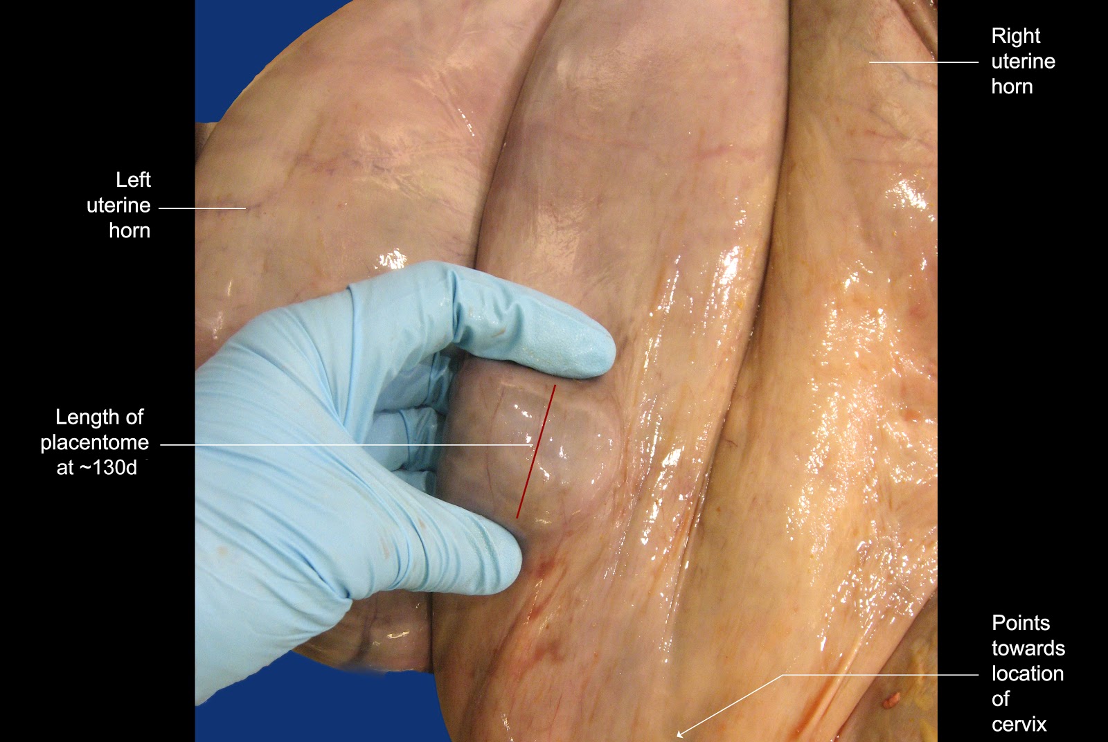
The image below shows how placentomes grow during pregnancy. Note that their growth is almost logarithmic in the last third of gestation.  The benchmarks discussed in the paragraph below are colored green in the image.


Image size: 1492x956px


Every operator will have her or his preferred method of remembering the size of placentomes at any given stage of pregnancy. The author provides the following guidelines for consideration. Again, these guidelines are used for palpation in the absence of ultrasonography. Measurements of structures using ultrasonography are highly accurate but when one takes tissue compression (during transrectal palpation) into account, mental impressions are not the same as as ultrasonographic measurements.

At 75 days, placentomes usually become discernible for the first time.

At 90 days (3 months) placentomes measure about 1 x 1.5 cm in size. In the author's rather unconventional mind, it is easily remembered because 1.5 is half of 3 but admittedly, this is barely rational.

At 120 days (4 months) placentomes measure approximately 2.5 x 1.5 cm in size. Added together, 2.5 and 1.5 equal 4 ; easy to memorize.

At 150 days (5 months) placentomes measure approximately 2 x 3 cm in size. Added together, 2 and 3 equal 5 ; easy to memorize.

After 5 months, the remarkable growth rate of placentomes become noticeable.

The dimensions of placentomes at 5 months of gestation are particularly useful to memorize because they are about that size in beef cattle during routine fall pregnancy diagnosis. Simply by spreading one's fingers slightly and applying pressure on the dorsal  surface of the uterus, then “raking’ in a cranial to caudal motion, a five month old pregnancy can be diagnosed easily. This procedure is so rapid and simple that hundreds beef cows can be examined in a few hours

At 6 months of gestation, the placentomes are approximately 6 cm long; again this is easy to memorize.

At term, the rapid growth of placentomes has increased their average length to approximately13 cm. Again, in the author's unconventional mind, this is remembered as “unlucky 13 at term".

Again, these benchmarks are 75 days, 3 months, 4 months, 5 months, 6 months and Term. Other durations are extrapolated between these benchmarks.
_____________________________

In the event that a fetus is aborted and one has to determine at what stage of gestation that occurred, many different guidelines are provided in the literature, some bordering on the ludicrous (small mouse, large rat without a tail etc). Instead, the following simple guidelines are offered. Again, one uses the dimensions of one's hand to make these determinations. A discussion follows the image.


Image size: 3046x2437px

The crown-rump length (CRL) of a two month old fetus extends across the palm of the author's hand as shown. At four months, the CRL extends between the tip of the author's middle finger and his wrist. Placing both hands, tip-to-tip as shown, covers the CRL of a six month old fetus. Finally, beyond the combined size of one's hands, the CRL increases to 80 cm at 8 months. Interestingly, these measurements apply to all ruminants and even horses. Incidentally, the CRL of an aborted 10 month old equine fetus is approximately 100cm.


Tuesday, November 22, 2016

Acardiac fetuses

 Acardiac fetuses (Amorphus globosus monsters)

Keywords: amorphous, globosus, (AG),(AGM), acardiac fetus (AF), bovine, placenta, monsters, karyotype, cattle

The essential nature of these fetuses is that they do not have hearts. Therefore the term amorphous globosus (AG) is less specific than acardiac fetus (AFs). AFs are placental parasites originating from embryos that do not develop into normal fetuses. Although the term "monster" is commonly applied to these fetuses because of their grotesque appearance, that term is melodramatic and archaic. Indeed, the term "monster" has been largely discarded in human medicine. The term anidian monster has also been used to describe an AF i.e. The cytological sex of a bovine anidian (amorphous) twin monster. HO Dunn et al. Cytogenetic and Genome Research, 1967. This term is seldom used and its etymology remains a mystery.

Should one consider amorphous globusus monsters (AFs) as fetuses? Merriam-Webster defines a fetus as: "An unborn or unhatched vertebrate especially after attaining the basic structural plan of its kind". On perusing descriptions of AFs it appears that they probably contain endoderm, ectoderm and mesoderm although these basic embryonic layers do not develop normally. If these three layers are considered to be a "basic structural plan" (could that not also be the DNA in a single cell?) then AFs should be referred to as fetuses.

The image below shows an acardiac quadruplet born together with three normal triplets females also shown in another LORI entry.


Image size: 2816 x 2070 px

It would be convenient to assume that this AF was also a female because the triplet females were not androgenized (freemartins). However, the genital system of the acardiac fetus had not developed, therefore the possibility of it having an XY karyotype could not be discounted (see below).

Below, an AF and a radiograph of that fetus (inset). The radiograph shows a small center of mineralization/ossification. The umbilical cord has broken free of the host placenta.


Image size: 947 x 719 px

A radiograph of a bilobed acardiac fetus with a highly mineralized/ossified focus.


Image size: 1200 x 1039 px

Acardiac fetuses are most common in cattle, followed by the small ruminants, then horses and rarely, humans. The reason that they occur more frequently in cattle than other animals is probably because placental fusion and anastomosis of blood supply is very common in cattle, also explaining the high frequency of freemartins in these animals. Without a blood supply joined to that of a viable fetus (see below) an acardiac fetus can not survive. 

In humans some play is made of the fact that the viable fetus is the "pump" fetus that supplies the abnormal fetus with blood. However, this is of course the situation with any acardiac fetus because a fetus without a heart must rely on blood flow from its placenta, provided by at least one other "pump" fetus in the uterus. Therefore "pump" fetuses occur in animals as well.

Acardiac fetuses occur most frequently in the form of amorphous masses with hair coats and umbilical cords. In unusual cases, they may be recognizable as fetuses with legs, faces, muzzles teeth, vulva lips and tails. Rarely, two acardiac monsters may accompany a normal fetus (triplets). 

As mentioned, AFs do not have hearts. Internally, they may have blood vessels, irregularly shaped bones, foci of mineralization, cartilage and fibrous tissue. In humans, AFs are usually monozygous and therefore the same sex but in cattle, the karyotypic sexes of the viable twin and the acardiac twin can be the same or different.  In addition, the karyotype of the AF can be normal or abnormal. The precise genetic abnormalities leading to acardiac development are not known.

It is considered unlikely that a female co-twin to an acardiac monster can be a freemartin (because of the lack of gross male gonads in the amorphous co-twin). However, it is possible that male gonadal tissue is present if the amorphous globusus co-twin is a male. Therefore, it is wise to examine the viable  female conceptus for freemartinism.

Selected references

Anwar. M.T. et al. 2009. A rare case of globosus amorphus in a goat. Can Vet.: 854–856.

Blaicher. W. et al. 2000 Acardiac twin pregnancy: associated with trisomy 2: Case report. Human reproduction. 15:474-475

del Rio, N.S et al. 2006. Observed frequency of monozygotic twinning in Holstein dairy cattle. Theriogenology. 66:1292-1299

Kamimura, S. 993 A globosus amorphus from an in vitro fertilized embryo transferred to a japanese black cow. Theriogenology 40:853-858

Pearson, L.K.et al Theriogenology Question of the Month. J. Am.Vet.Med.Assn 238:1261-1263

Roberts, S.J. 1986. Veterinary obstetrics and genital diseases. Gestation period, pp 79-81. Published by the author, S.J. Roberts.

Weber J et al. 2017. Facets of Clinical Appearance and Aetiology in an Unusual Bovine Amorphus Globosus. Anat. Histol. Embryol. 46:502–506


Friday, November 4, 2016

Embryonic/fetal membrane slipping

Keywords: membrane, slip, fetus, embryo, diagnosis, pregnancy, ruminant, bovine, ovine, caprine, staging.

Membrane slip is commonly used to diagnose pregnancy in cattle, especially in dairy cattle where early pregnancy diagnosis is important. Membrane slip manifests itself when gentle pressure is applied to a fold of the uterine wall. If a conceptus is present, its free allantois or allantochorion or even the free chorion itself, slips away from the constricted section of the uterus. This creates a distinct, palpable "click" between the operator's fingers. 

The composite image below shows how a membrane slip is generated. In image A, the uterus is grasped between the thumb and any finger, then the digits are rolled relative to one another until a firm clicking sensation is detected. The operators digits are not moved off the uterus at any time during this operation. Using an opened, pregnant uterus in image B, one can see how the edge of embryonic/fetal membrane (yellow arrows) is pinched gently between the operator's thumb and finger. As these digits are rolled relative to one another (decreasing the interposing angle between the thumb and finger) the membrane escapes the operator's grip (red and yellow arrows in image C) generating the clicking sensation.


Image size: 1024 x 768px.

In several studies, membrane slipping has been shown to be harmless to the conceptus.

As reviewed elsewhere in LORI, during early gestation, the allantois is not yet attached to the chorion. However, these two membranes gradually become inseparable and collectively, are known as the allantochorion or less commonly, the chorioallantois. Therefore in early gestation, it is either the allantois or chorion or allantochorion that is involved in membrane slipping; a fairly trivial point.

Although the production of pregnancy specific proteins, RNA segments and Interferon Tau have been developed for early pregnancy detection in cattle, ultrasonography is currently accepted as the gold standard for comparison of pregnancy diagnostic methods in cattle; it is both highly specific and sensitive after 28 days of gestation and especially valuable for the diagnosis of twins. However, membrane slipping continues to be a valuable diagnostic tool for routine pregnancy diagnosis. Membrane slipping is also a useful tool for the detection of early pregnancy in small ruminants and wild ruminants when laparotomy is performed for any reason. It cannot be used in new world camelids or equids because of the diffuse placental attachment in those animals.

Comment on the use of the word "fetal" in fetal membrane slip, still a widely accepted term in animal science:

It has been stated that the embryonic period in cattle extends from fertilization to the completion of differentiation at about 42 days (Committee on Reproductive Nomenclature, 1972. Recommendations for standardizing bovine reproductive terms. Cornell Vet 62: 216–237). However it is arguable that differentiation continues throughout pregnancy or even postnatal life and therefore, that the distinction between an embryo and fetus is arbitrary. Even in humans, where an embryo makes its sudden and completely artificial transition into fetal life at 8 weeks, this terminology is under review. Therefore, in this author's opinion, it is erroneous to use fetal membrane slip as a general diagnostic term in cattle, especially dairy cattle. This is because fetal membrane slip is most often used as a diagnostic tool before 42 days, when the conceptus is still formally classified as an embryo. This may be a trivial matter (and in the greater scheme of things, probably is) but like the term embryotomy it should probably be discarded in favor of a more global and accurate term. In that light, the author suggests that instead of the term fetal membrane slip, consideration be given instead, to using the term membrane slip throughout pregnancy.

Selected references:

Ioannidis, J. 2016. Circulating miRNA signatures of early pregnancy in cattle. BMC Genomics. 17: 184

Nation, D.P. et al. 2003. Accuracy of bovine pregnancy detection using transrectal ultrasonography at 28 to 35 days after insemination. Australian Vet. J. 81: 63–65

Preeti Rawat et al. 2016. Identification of potential protein biomarkers for early detection of pregnancy in cow urine using 2D DIGE and label free quantitation. Clin Proteomics. 13: 15.

Romano, J.E. 2006. Early pregnancy diagnosis by transrectal ultrasonography in dairy cattle.
Theriogenology. 66:1034–1041


Wednesday, March 2, 2016

An involuting uterus, late postpartum

Keywords: uterus, vagina, bovine, corpus luteum, postpartum, cervix

The uterus of a cow that had involuted partially after calving. She had also ovulated at least once after calving.


Image size: 1308 x 972px

The presence of a corpus luteum (CL) in the left ovary indicates that she had ovulated recently. 

Some may ask: Why is it that a cow would ovulate before her uterus is fully involuted and ready to accept a new pregnancy? The answer is that this cow's calf was removed shortly after birth and was not allowed to suckle i.e. this tract was from a dairy cow, a product of human manipulation. Dairy cows are not subjected to the negative effect of sucking on postpartum anestrus. By contrast, beef cows are suckled and do not ovulate for extended periods after calving (depending on breed and nutrition) usually well after the time that the uterus has taken to involute completely. 

In the right ovary of this cow, a small follicles has formed a layer of luteal tissue within the follicle, a sign of low level stimulation with luteinizing hormone (LH). Although it is smaller than most cystic follicles it is essentially just that; a luteinized cystic follicle. It is possible that this cystic follicle could have persisted beyond the lifespan of the CL in the left ovary, causing the cow to have a delayed return to estrus (as is often the case). However it is also possible that it was of a similar age to the CL, failing to mature and ovulate as a twin ovulation. 

Finally, note that there is no shortage of small tertiary follicles in these ovaries, waiting to participate in postpartum estrous cycles. This is because the production of follicle stimulating hormone (FSH) recovers far sooner that LH production after a calf is born.

The caruncles in this specimen are small and almost completely involuted yet discoloration in the centers on the caruncles suggests that their involution is not complete. The endometrium between caruncles is sloughed soon after calving but is restored by 10 to 12 days postpartum. The caruncles by contrast, have only sloughed tissue to the level of their capillary beds by that time and it is only by about 35 days  postpartum that caruncles are completely restored with new epithelium. Interestingly, caruncles visible throughout the life of the cow. In fact they are even evident when females calves are developing in utero.

Note the short uterine body i.e. how quickly the horns divide as an object ascends into the tract. This is important for several reasons. First, if one is to preferentially inseminate in one horn or another, the pipette must be directed accordingly, as soon as the internal cervical os has been reached, A similar situation is encountered when one has to collect embryos from one horn or another. Finally, because of the virtual absence of a uterine body, bovine fetuses are highly likely to be in longitudinal presentation at calving. By contrast, mares have long uterine bodies making transverse presentation more likely than in cows (although thankfully, transverse presentations are still unusual in mares). Also, no thought is given to horn selection in mares during routine insemination or embryo flushing.

Note the cervical "rings". Apart from the ring that surrounds the external cervical os, these structures are more akin to crescents than complete rings. One must negotiate these rings with an insemination pipettes during artificial insemination (AI). In mares, these rings do not exist and entry into the uterus is simple. In cattle by contrast, the cervix must be grasped per rectum and the tip of the A.I pipette placed in the fornix of the vagina. Then the external cervical os is then manipulated over the instrument while the instrument itself is eased through the cervical canal. The same situation is encountered when one flushes embryos from the uterus.

Interestingly, the word fornix is derived from the Latin word for archway. In homo sapiens, standing erect, this is perhaps appropriate. In domestic animals however, the arches (around the cervix) have fallen on their sides!

Selected reference:

The mind of an aging theriogenologist.

Amniotic plaques

Keywords: bovine, artiodactyls, amnion, amnionic, amniotic plaques, pregnancy

Amniotic or amnionic? Both words have the same meaning and are widely used in the scientific literature. However, a search on Google Scholar revealed 440,000 hits for amniotic and about one tenth of that number (only 4,770 hits) for amnionic. Therefore the term amniotic is more widely used than amnionic and accordingly, has been adopted throughout this LORI entry. Incidentally the etymology of the term amnion is not clear. Some references define it simply as having originated from the same word in Latin meaning "the membrane over a lamb's head"

The image below shows the amnion and fetus from a 95 to 100 day old pregnancy. The allantois and chorion have been removed for clarity.  Click on image for larger version.

The whitish-grey plaques distributed towards the center of the amnion are normal findings yet their presence is an enigma; they are amniotic plaques. Because of their histologic resemblance to skin, they have also been referred to as "callusoids" (as in a callous) or "verrucoids" ( < L. verrucosus; resembling a wart).


Amniotic plaques are most obvious in artiodactyls (cloven hoofed animals) especially cattle but they are in fact found in the majority of mammals, including cetaceans, humans and even sloths (a fact of major importance to sloths alone).  In some cases, they are barely visible to the naked eye. Indeed, among domestic animals they also occur in the horse; our only perissodactyl. Yet there again, they are very small and often overlooked. Although amniotic plaques are more common in abnormal pregnancies in humans, they occur in normal pregnancies as well. Interestingly, in abnormal bovine pregnancies arising from cloned embryos, they are better developed than otherwise and are clearly visible on ultrasonography; something not usually possible in normal bovine pregnancies.

Amniotic plaques are found on the inside of the amnion and this should be obvious on second thought because close observation shows them to be present on the outside of the umbilical cord, which is of course, inside the amnion. Nevertheless, the amnion can be so flaccid as to give the impression that the plaques are on the outside of the membrane. That phenomenon is seen below; the fetus still within the amnion.


Image size: 1200x793px

Histology shows amniotic plaques to have a remarkable resemblance to skin with perhaps 30 layers of tissue, considerably thicker than the surrounding amnion which may be only four or five cells thick.  Like skin, they also have layers such as stratum basale, stratum spinosum, stratum granulosum and stratum corneum.

Light microscopy as well as scanning and transmission EM have been used to study amniotic plaques in cattle, humans and some animals (deer and sheep). It has also been shown that the amnion produces low molecular weight protein throughout gestation; yet the function of amniotic plaques is still unknown. Even more intriguing is the fact that they are temporary structures, first visible in cattle at about three months of gestation becoming well developed by five months or so, then gradually disappearing and being completely absent at term. In other animals they are temporary structures as well.

Note: The General Assembly of the World Association of Veterinary Anatomists (2006) contrived the term “bractreolae amnioticae”  as an internationally acceptable synonym for the English term amniotic plaques. The general response to this term amoung English speaking colleagues is either a quizzical look or a vacant stare.

Selected references:

Ginther, O.J. 1992 Reproductive biology of the mare and applied
aspects, 2nd ed. Cross Plains, WI: Equiservices. p.379

Kohan-Ghadr, H.R. et al. 2008. Ultrasonographic and histological characterization of the placenta of somatic nuclear transfer-derived pregnancies in dairy cattle. Theriogenology: 69:218-230

Lui, K.H. et al. 1994. Ultrastructure and protein synthesis of bovine amnion. Animal Reprod Sci. 35: 41-55

Mossmann, H.W. 1987. Vertebrate fetal membranes. Rutgers University Press. ISBN: 0-8135-1132-1 pp158-159.

Roberts, S.J. 1971 Veterinary Obstetrics and Genital Diseases. Ithaca, NY, Published by the author, p 43

Schlafer, D.H. et al, 2000. The bovine placenta before and after birth: placental development and function in health and disease. Anim. Reprod, Sci. 60-61:145-160.

Sinha, A. A. et al. 1970. Ultrastructure of the amnion and amniotic plaques of the white-tailed deer. Am.J.Anatomy. 127: 369–395

Wislocki, G.B. 1941. The placentation of an antelope (Rhynchotragus kirkii Nyikae Heller).
The Anat. Record. 81: 221–241