BOVINE IMAGES

How to use these images


IMPORTANT: To see an enlarged image, click on any image you see in LORI. Then, RIGHT click on the enlarged image to save it at its full size.


Showing posts with label calving. Show all posts

Friday, April 21, 2017

Late gestation pregnancy and placentation

Keywords: bovine, calving, partus, parturition, placenta, caruncles, placentomes, cotyledons, cervix

These images show some of the characteristics of a pregnant uterus removed from a Simmental cow that died suddenly during the early stages of calving. The cause of death was not determined.

As shown below, the calf was located in the right uterine horn. In that regard, it is useful to remind readers that the uterine horn occupied by the fetus is of little significance during cesarean sections. This is because the so called "non-pregnant" horn contains such a small volume of fetal fluid that it is deflected towards the midline by the horn that contains the calf. Therefore (in a normal pregnancy) albeit left or right, the "pregnant" horn is readily accessible from a normal left flank approach.


Image size: 2888 x 1585 px 

In the image above, arrow A indicates that if one were to raise the right uterine horn and inspect the mesometrium (arrow B) the uterine artery would be visible. From about 4 months of gestation, the volume of blood flow in this artery excedes the ability of its thin wall to constrain blood flow in a linear fashion. In bovine pregnancies, this results in turbulent blood flow, reminiscent of air flowing through a "wind sock". In this video, the author illustrates turbulent air flow that is akin to blood flow vibration felt during palpation. This phenomenon is known as fremitus (< Latin. "To murmur"). Fremitus appears later in the vessel supplying the "non-pregnant" horn than that supplying the "pregnant" horn. In both vessels however, it ceases shortly after calving. Therefore it can still be present after calving and in the strictest sense, cannot be regarded as an infallible indicator of pregnancy.

Interestingly, fremitus is not detectable in pregnant mares.

As shown below, the corpus luteum (CL) of pregnancy (ringed in green) was still present in this specimen, shortly before calving would have occurred. This is normal.


Image size: 1081 x 710 px

Although the CL of pregnancy is still present at term, most cows remain pregnant even if total ovariectomies are performed within the last 30 days of gestation. Indeed, some cows becomes independent of CL progesterone secretion as early as 6 months of gestation. After ovariectomy however, pregnancies do not end normally. Although pregnancies are maintained to term, cervical dilation, dystocia and retained fetal membranes are common. This is because the corpus luteum is a also a source of relaxin, prostaglandins E1 & E2 and even oxytocin. It is therefore, essential for normal parturition.

Soon (12 to 16 hours) after substantial uterine contractions begin, the cervix relaxes rapidly. This relaxation is largely under the effect of PGE2 but as mentioned earlier, other hormones are also involved.

The initial phase of cervical dilation is largely passive i.e. independent of the dividing force of the fetal head and forelimbs. However, the effect of the fetus can not be excluded completely because the uterus is pushing it towards the cervix, even during the early stages of cervical dilation. Within 8 to 10 hours in cows and somewhat longer in heifers, the cervical canal dilates to a diameter of 12 to 20 centimetres i.e. big enough to accommodate the head of the foetus.


Image size: 1000 x 772 px

In the specimen discussed here, passive dilation was incomplete yet almost sufficient to accommodate a human hand (about 12 cm wide, including the thumb).


Image size: 3111 x 1729 px

Normally, in pre-term pregnant cows, even in autolysed specimens, this would be impossible. Therefore had this cow lived, the second (expulsive) stage of calving would have been imminent.

The image below confirms the fact that the fetus had not yet entered the birth canal i.e. active dilation of the cervix and second stage parturition had not yet begun.


Image size: 2138 x 1056 px

The following image shows the fetal membranes and the calf, removed from the uterus.  The normal number of placentomes in bovine pregnancies varies between 75 and 120, the largest measuring 12 to 14 cm in length. Also, as reviewed elsewhere, amnionic fluid volume varies between 2 and 8 liters and allantoic fluid, between 4 and 15 liters. This pregnancy appeared to be normal in those respects.


Image size: 3456 x 2130 px

Placentation in ruminants consists of numerous complexes. Hence the term "multiplex placentation" as apposed to "diffuse, discoidal, zonary" etc.  The two parts of each complex are of course, a caruncle on the maternal side and a cotyledon on the fetal side. Together, they form a placentome. A placentome is shown here:


Image size: 1200 x 809 px

As illustrated elsewhere in LORI, the site and number of caruncles in a cow has already been established when the cow herself was a fetus!

Note how the fetal villi are sliding out of the maternal crypts, a process that is impossible if  placental maturation (flattening of maternal epithelium and loosening of fetal-maternal contact) has not occurred during the last few days of gestation. Incomplete placental maturation is a cause of retained placenta. Placental maturation is impaired if the fetus is born prematurely or the diet of the cow is deficient in selenium and vitamin A, nutrients that are essential for epithelial function. This explains in part why retained placenta is common when selenium and vitamin A are deficient in the diet of pregnant cattle.

Tuesday, January 19, 2016

Prolapsed uterus 

Keywords: drawing, Trengrove, bovine, calving, accident, prolapse

Author: Dr Ronald Trengrove, modified by Dr Rob Lofstedt.

This drawing, by Dr Ronald Trengrove, a colleague and ex-teacher at Onderstepoort Veterinary institute, RSA, provides an excellent explanation of uterine prolapse in a cow. It is with his permission that it was provide for LORI. Dr Trengrove died in 2015.

When a cow has calved a large fetal-placental load (males, twins, hydrops etc) or secondary intertia follows dystocia, the myometrium lacks tone. The same occurs in cases of hypocalcemia. In both situations, the uterus may move caudally under the effect of abdominal straining or gravity (when the cow lies in a pasture with her hind quarters downhill). As shown in figure a, involution occurs first, then, as the double wall of the uterus enters the birth canal (figure b), its bulk stimulates abdominal straining, expelling the uterus and completing the prolapse (figure c).


Image size 900 x 805px.

In most cases, neither the mesometrium nor its blood vessels rupture during prolapse. After cleaning and repair, the uterus can be returned to its normal situation by elevation, compression and pressure in a cranial direction. The value of topical osmotic agents such as magnesium sulphate and sugar is debatable. Once the uterus is in situ, a Buhner suture as used for vaginal prolapse can be applied to the vulva lips and both calcium and oxytocin can be administered to restore myometrial tone.

In the event that the uterus is severely damaged and cannot be replaced, a drastic salvage procedure was described by Dr Trengrove. The author has never attempted this and has not seen it described elsewhere. Using epidural anesthesia, the cow is made recumbent and the prolapsed uterus is hoisted over a plank (a). After using needle puncture or ultrasound to exclude the presence of the bladder, a major tourniquet (b) using Buhner tape is placed adjacent to the vulva lips. Then a split ligature of umbilical tape is placed more distally (c) and the uterus is transected along the length of the plank as shown.  The stump is then replaced. Presumably analgesics and antibiotics are used to manage the cow so that she is able to recover and wean her calf.


Image size 469 x 446px

Monday, August 5, 2013

Routine assistance during calving.


Keywords: obstetrics, calving, bovine, cow, twins, presentation, position, posture


Image size: 1568 x 1040 px

This series of images illustrates standard procedure for attending to a calving. In this case, the dam was a pluriparous Hereford cow.

In most cases, pluriparous cows give birth within one hour. Up to three hours may elapse before calving is complete in a heifer. After those time frames, assistance should be considered. If however, one is present at the onset of calving, gentle and quiet intervention may be beneficial. In animals not accustomed to humans, unnecessary intervention may delay calving and should be avoided.

In this image (number one) the amnion has just begun to show at the vulva lips. This means that the allantochorion (chorioallantois) has ruptured already, releasing allantoic fluid.

Routine assistance during calving. 2.


Image size: 1912 x 1312 px

In this image (number two) the amnion is still intact and two forelimbs and the nose of the calf has been presented at the vulvar lips. Normally, presented cranially and longitudinally. The position of the calf is usually dorso-left ilial or dorso right- ilial, moving into dorsosacral quite quickly. Normal posture for the calf is with its head resting upon its two forelimbs. If calving appears to be progressing slowly, it may be beneficial to rupture the amnion manually and assess presentation, position, and posture before dystocia becomes established.

Routine assistance during calving. 3.


Image size: 18988 x 1333 px

In this image (number three) the operator is scrubbing his hands and arms in preparation for intervention. It is important to exercise good hygiene if assistance is to be given during calving. In this case an iodine-based scrub is being used but for practical purposes, there are physical cleansing with ordinary soap and water is usually sufficient. The use of gloves is not recommended; traction is compromised and handling of the calf and fetal membranes is complicated through the use of gloves.

Waterproof, washable calving suits are available but may be uncomfortable in hot weather.  Otherwise, clothing should be discarded and washed.

Routine assistance during calving. 4.


Image size: 1961 x 1251 px

In this image (number four) the operator has ruptured the amnion manually and some amnionic fluid has escaped. Normally there are about 8 L of amniotic fluid and about twice that amount of allantoic fluid.

Routine assistance during calving. 5.


Image size: 1707 x 1316 px

In this image (number five) the operator has established that the head and forelimbs presented at the vulvar lips belonged to the same calf! It is possible in some cases for twins to be presented simultaneously; this possibility should be eliminated before any traction is applied to the head or limbs; exacerbating dystocia.

Routine assistance during calving. 6.


Image size: 1928 x 1312 px

In this image (number six) traction is being applied to both forelimbs and the head is following spontaneously. In cases of dystocia, calving chains and perhaps even a head snare would have been applied to increase purchase on the calf. It is sometimes difficult to decide exactly how much traction should be applied and when too much traction is necessary to deliver the calf per vagina (necessitating a cesarean section). In general, two average sized adults should be able to deliver a calf per vagina if its presentation, posture, and position are normal. If it is obvious that the fetus is relatively or actually oversized, as is often the case with beef cattle, one should defer to a cesarean section as soon as possible.

Routine assistance during calving. 7.


Image size: 1950 x 1307 px

In this image (number seven) the elbows of the calf had been grasped and the calf has been rotated through about 120° so that the widest measurement of the fetal pelvis (across the heads of the femoral trocanters) lies dorsal ventral in the maternal pelvis. This helps to prevent hip lock in the maternal pelvis.

Routine assistance during calving. 8.


Image size: 1917 x 1298 px

In this image (number eight) mucus and other debris is being removed from the calf's mouth. If suction is available, it should be applied now. Some ranchers hang calves vertically for a few minutes at this time or even swing them around centrifugally in an effort to expel the mucus in the upper respiratory tract. However this may put excessive pressure on the diaphragm and compromise oxygenation therefore is generally not advisable.  Oxygen can be administered by face mask if available. The value of buffers (NaBicarbonate and TRIS) as well as analeptics such as doxapram hcl are questionable.

At this time, it is also appropriate to check for the presence of a cleft palette in the calf.

Routine assistance during calving. 9.


Image size: 1963 x 1312 px

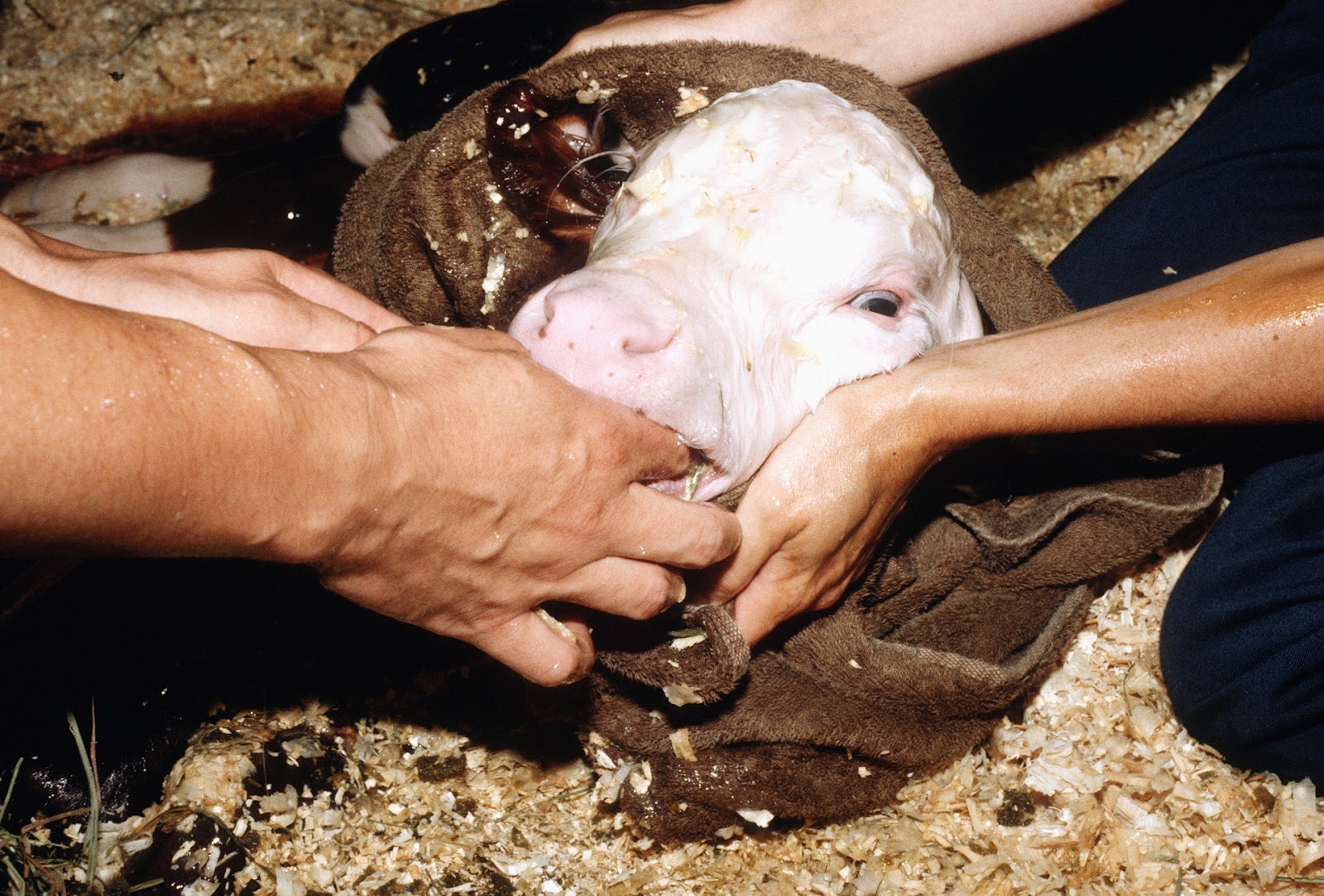
In this image (number nine) the calf is being rubbed vigorously in an attempt to vitalize it; mimicking the vigorous licking of the calf by the dam that normally occurs at this time. One should determine the gender of the calf and also ensure that no umbilical or inguinal herniation is present. The umbilical cord should be disinfected with 0.5% chlorhexidine. Disinfection should be repeated 12 to 24 hours later.

Routine assistance during calving. 10.


Image size: 1927 x 1298 px

In this image (number 10) a helper is continuing to stimulate the calf, close to the dam's head, allowing some fetal-maternal bonding to occur. Excessive helping or interference may compromise fetal-maternal bonding, especially in animals not habituated to humans. At this time the operator is examining the reproductive tract for any signs of damage that may have occurred during delivery and importantly is also examining the uterus for the presence of a second calf.  Intra-uterine antibiotics or disinfectants should not be administered routinely after calving. Their value is questionable; indeed this practice has also been associated with an increased calving to conception interval.

Routine assistance during calving. 11.


Image size: 1878 x 1312 px

In this image (number 11) a second calf was indeed present. It was delivered in the same manner as the first calf and the uterus was again investigated for the presence of a third calf which was not present. At this time, the dam should be allowed to bond with her calves with minimal interference from humans.

Terminology: 

Uniparous, monotocous, polytocous, multiparous, primiparous, pluriparous, polytocous and. What do these terms mean?

Monotocous often carries the same meaning as uniparous but is  mainly used in zoology and veterinary medicine. Although uniparous is often used instead of monotocous in human medicine, that is essentially incorrect because uniparous can also refer to an animal that has only one offspring in its life. Therefore, the term monotocous should be used for any animal (including humans) that usually produces a single offspring in any pregnancy. 

Primiparous animals those that are giving birth for the first time 

Multiparous animals are those that usually have had more than one offspring in subsequent pregnancies.

Polytocous are those that USUALLY give birth to more that one offspring per pregnancy.

For example: A  heifer is usually a monotocous animal but can occasionally be polytocous, producing twins and rarely triplets even as primiparous animal. After the birth of its twins or even triplets it is still referred to as a primiparous animal. After its second, third or Nth pregnancies, it a pluriparous animal but still referred to as monotocous! Essentially then "parity" refers to the number of pregnancies, while tocous refers to the number of fetuses. One final thing: -tocos is the noun, -tocous, the adjective!