BOVINE IMAGES

How to use these images


IMPORTANT: To see an enlarged image, click on any image you see in LORI. Then, RIGHT click on the enlarged image to save it at its full size.


Showing posts sorted by relevance for query pregnancy. Sort by date Show all posts

Friday, November 4, 2016

Embryonic/fetal membrane slipping

Keywords: membrane, slip, fetus, embryo, diagnosis, pregnancy, ruminant, bovine, ovine, caprine, staging.

Membrane slip is commonly used to diagnose pregnancy in cattle, especially in dairy cattle where early pregnancy diagnosis is important. Membrane slip manifests itself when gentle pressure is applied to a fold of the uterine wall. If a conceptus is present, its free allantois or allantochorion or even the free chorion itself, slips away from the constricted section of the uterus. This creates a distinct, palpable "click" between the operator's fingers. 

The composite image below shows how a membrane slip is generated. In image A, the uterus is grasped between the thumb and any finger, then the digits are rolled relative to one another until a firm clicking sensation is detected. The operators digits are not moved off the uterus at any time during this operation. Using an opened, pregnant uterus in image B, one can see how the edge of embryonic/fetal membrane (yellow arrows) is pinched gently between the operator's thumb and finger. As these digits are rolled relative to one another (decreasing the interposing angle between the thumb and finger) the membrane escapes the operator's grip (red and yellow arrows in image C) generating the clicking sensation.


Image size: 1024 x 768px.

In several studies, membrane slipping has been shown to be harmless to the conceptus.

As reviewed elsewhere in LORI, during early gestation, the allantois is not yet attached to the chorion. However, these two membranes gradually become inseparable and collectively, are known as the allantochorion or less commonly, the chorioallantois. Therefore in early gestation, it is either the allantois or chorion or allantochorion that is involved in membrane slipping; a fairly trivial point.

Although the production of pregnancy specific proteins, RNA segments and Interferon Tau have been developed for early pregnancy detection in cattle, ultrasonography is currently accepted as the gold standard for comparison of pregnancy diagnostic methods in cattle; it is both highly specific and sensitive after 28 days of gestation and especially valuable for the diagnosis of twins. However, membrane slipping continues to be a valuable diagnostic tool for routine pregnancy diagnosis. Membrane slipping is also a useful tool for the detection of early pregnancy in small ruminants and wild ruminants when laparotomy is performed for any reason. It cannot be used in new world camelids or equids because of the diffuse placental attachment in those animals.

Comment on the use of the word "fetal" in fetal membrane slip, still a widely accepted term in animal science:

It has been stated that the embryonic period in cattle extends from fertilization to the completion of differentiation at about 42 days (Committee on Reproductive Nomenclature, 1972. Recommendations for standardizing bovine reproductive terms. Cornell Vet 62: 216–237). However it is arguable that differentiation continues throughout pregnancy or even postnatal life and therefore, that the distinction between an embryo and fetus is arbitrary. Even in humans, where an embryo makes its sudden and completely artificial transition into fetal life at 8 weeks, this terminology is under review. Therefore, in this author's opinion, it is erroneous to use fetal membrane slip as a general diagnostic term in cattle, especially dairy cattle. This is because fetal membrane slip is most often used as a diagnostic tool before 42 days, when the conceptus is still formally classified as an embryo. This may be a trivial matter (and in the greater scheme of things, probably is) but like the term embryotomy it should probably be discarded in favor of a more global and accurate term. In that light, the author suggests that instead of the term fetal membrane slip, consideration be given instead, to using the term membrane slip throughout pregnancy.

Selected references:

Ioannidis, J. 2016. Circulating miRNA signatures of early pregnancy in cattle. BMC Genomics. 17: 184

Nation, D.P. et al. 2003. Accuracy of bovine pregnancy detection using transrectal ultrasonography at 28 to 35 days after insemination. Australian Vet. J. 81: 63–65

Preeti Rawat et al. 2016. Identification of potential protein biomarkers for early detection of pregnancy in cow urine using 2D DIGE and label free quantitation. Clin Proteomics. 13: 15.

Romano, J.E. 2006. Early pregnancy diagnosis by transrectal ultrasonography in dairy cattle.
Theriogenology. 66:1034–1041


Saturday, December 3, 2016


Guidelines for aging bovine pregnancies.

Keywords: hand, measurement, abortion, pregnancy, diagnosis, bovine, placentomes, crown-rump

It is not the author's intention to foist a personal bias onto colleagues; rather, to provide another system for students to entertain. Over the years, those who work with cattle develop methods that work for each of us; differing from one operator to the next. The methods mentioned here arise from the author's experience.

In this entry, the terms "pregnancy" and "gestation" are used interchangeably where appropriate.

In the absence of ultrasonography, an excellent way of improving aging accuracy is to ask a farmer not to provide the breeding date before an attempt is made at aging the pregnancy. It should be possible to  determine the age of a pregnancy within at least, half of the duration of an estrous cycle (~10.5 days). In this manner, the farmer can determine the "dry-off" date without losing production or shortening the dry period unnecessarily. Between 42 and perhaps 90 days of gestation, it is frequently possible to determine the duration of gestation within four or five days but as the duration of gestation lengthens, one's accuracy decreases. Fortunately, the first third of gestation is usually the most critical for dairy farmers; not only must pregnancy be confirmed with certainty but in cases of re-breeding, it is often necessary to determine which insemination has accounted for the pregnancy.

There is arguably, only one cardinal sign of pregnancy i.e. fetal membrane slip. Enlargement of the uterus can be due many things other than pregnancy. Fremitus of the uterine artery can persist after calving or abortion and placentomes are palpable in those cases as well. Therefore, only after a fetal membrane slip has been detected, should one proceed to estimate the age of a pregnancy.

It is incorrect to refer to a "pregnant" horn because the fetal membranes on the side of ovulation occupy the contralateral horn by 18 to 20 days of gestation. Nevertheless, this convention persists and for convenience, is also used in this LORI entry.

In the image below (its detail only becoming evident when enlarged) the author's hand is shown as though it has grasped the pregnant horn in each case. The diameter of the uterus is represented by the area between the thumb and forefinger. Measurements for average male hands (the author's hands) and average female hands are shown at lower right in the image. Readers are encouraged to become familiar with the size of their own hands.

It is important to realize that a heifer's uterus is perhaps ten to twenty percent smaller than that of a multiparous cow at the same stage of pregnancy. Appropriate accommodation must be made for this fact when estimating the duration of  gestation in a heifer. The growing uteruses in this image approximate those of a cow, not a heifer.


Image size: 2849x 2020px

The dimensions of a uterus are not palpable with great accuracy after four months of gestation because its cranial margin has grown beyond the reach of most operators. In fact, by five months of gestation, it is virtually impossible to palpate the cranial margin of the uterus. At five months, the fetus itself has sunk so low within the abdomen that it too, may not be palpable. After six or seven months, the fetus becomes large enough to palpate once again.

The author has found it to be of great value to acquaint himself with the measurements of his hands, not only for estimating the duration of pregnancy or the age of aborted fetuses but for many other estimations too. Once one becomes acquainted with the dimensions of one's hands, their accuracy as measuring instruments are remarkable.

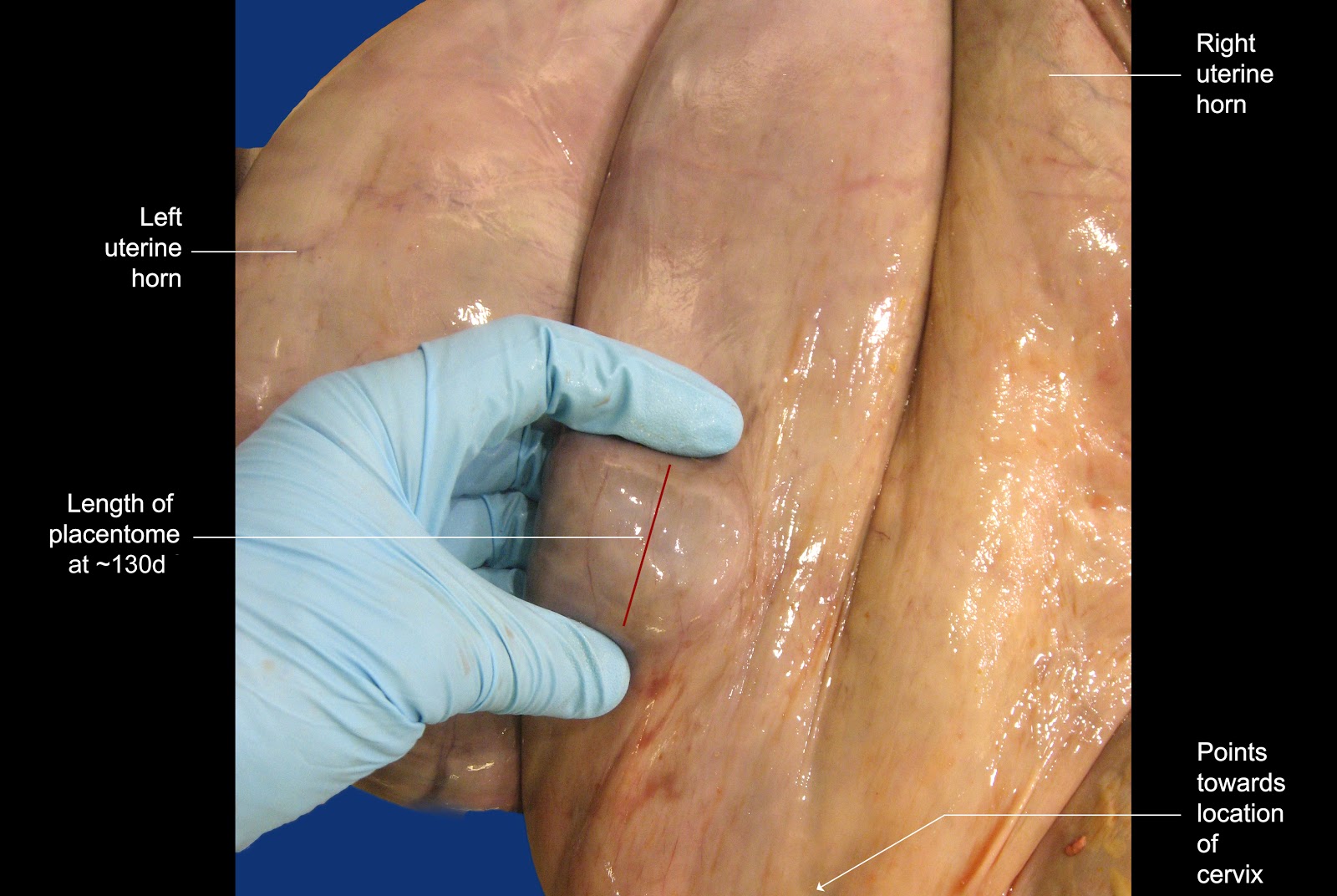
The image below shows the general area in which placentomes are palpated. In essence, one only uses the placentomes close to the cervix to make estimations of the size of these structures. If one palpates more cranial than this, it is likely that the duration of the pregnancy will be over estimated because placentomes generally become larger as one's hand moves cranially over the “pregnant” horn.


Image size: 3660x 2112px

Although the third image shows a placentome conveniently placed between thumb and forefinger for a measurement, this is not always possible in practice. More often, to gain an impression of their average size, several placentomes are manipulated between one's fingers while pressing down within the rectum.


Image size: 3152x 2112px

The image below shows how placentomes grow during pregnancy. Note that their growth is almost logarithmic in the last third of gestation.  The benchmarks discussed in the paragraph below are colored green in the image.


Image size: 1492x956px


Every operator will have her or his preferred method of remembering the size of placentomes at any given stage of pregnancy. The author provides the following guidelines for consideration. Again, these guidelines are used for palpation in the absence of ultrasonography. Measurements of structures using ultrasonography are highly accurate but when one takes tissue compression (during transrectal palpation) into account, mental impressions are not the same as as ultrasonographic measurements.

At 75 days, placentomes usually become discernible for the first time.

At 90 days (3 months) placentomes measure about 1 x 1.5 cm in size. In the author's rather unconventional mind, it is easily remembered because 1.5 is half of 3 but admittedly, this is barely rational.

At 120 days (4 months) placentomes measure approximately 2.5 x 1.5 cm in size. Added together, 2.5 and 1.5 equal 4 ; easy to memorize.

At 150 days (5 months) placentomes measure approximately 2 x 3 cm in size. Added together, 2 and 3 equal 5 ; easy to memorize.

After 5 months, the remarkable growth rate of placentomes become noticeable.

The dimensions of placentomes at 5 months of gestation are particularly useful to memorize because they are about that size in beef cattle during routine fall pregnancy diagnosis. Simply by spreading one's fingers slightly and applying pressure on the dorsal  surface of the uterus, then “raking’ in a cranial to caudal motion, a five month old pregnancy can be diagnosed easily. This procedure is so rapid and simple that hundreds beef cows can be examined in a few hours

At 6 months of gestation, the placentomes are approximately 6 cm long; again this is easy to memorize.

At term, the rapid growth of placentomes has increased their average length to approximately13 cm. Again, in the author's unconventional mind, this is remembered as “unlucky 13 at term".

Again, these benchmarks are 75 days, 3 months, 4 months, 5 months, 6 months and Term. Other durations are extrapolated between these benchmarks.
_____________________________

In the event that a fetus is aborted and one has to determine at what stage of gestation that occurred, many different guidelines are provided in the literature, some bordering on the ludicrous (small mouse, large rat without a tail etc). Instead, the following simple guidelines are offered. Again, one uses the dimensions of one's hand to make these determinations. A discussion follows the image.


Image size: 3046x2437px

The crown-rump length (CRL) of a two month old fetus extends across the palm of the author's hand as shown. At four months, the CRL extends between the tip of the author's middle finger and his wrist. Placing both hands, tip-to-tip as shown, covers the CRL of a six month old fetus. Finally, beyond the combined size of one's hands, the CRL increases to 80 cm at 8 months. Interestingly, these measurements apply to all ruminants and even horses. Incidentally, the CRL of an aborted 10 month old equine fetus is approximately 100cm.


Friday, April 21, 2017

Late gestation pregnancy and placentation

Keywords: bovine, calving, partus, parturition, placenta, caruncles, placentomes, cotyledons, cervix

These images show some of the characteristics of a pregnant uterus removed from a Simmental cow that died suddenly during the early stages of calving. The cause of death was not determined.

As shown below, the calf was located in the right uterine horn. In that regard, it is useful to remind readers that the uterine horn occupied by the fetus is of little significance during cesarean sections. This is because the so called "non-pregnant" horn contains such a small volume of fetal fluid that it is deflected towards the midline by the horn that contains the calf. Therefore (in a normal pregnancy) albeit left or right, the "pregnant" horn is readily accessible from a normal left flank approach.


Image size: 2888 x 1585 px 

In the image above, arrow A indicates that if one were to raise the right uterine horn and inspect the mesometrium (arrow B) the uterine artery would be visible. From about 4 months of gestation, the volume of blood flow in this artery excedes the ability of its thin wall to constrain blood flow in a linear fashion. In bovine pregnancies, this results in turbulent blood flow, reminiscent of air flowing through a "wind sock". In this video, the author illustrates turbulent air flow that is akin to blood flow vibration felt during palpation. This phenomenon is known as fremitus (< Latin. "To murmur"). Fremitus appears later in the vessel supplying the "non-pregnant" horn than that supplying the "pregnant" horn. In both vessels however, it ceases shortly after calving. Therefore it can still be present after calving and in the strictest sense, cannot be regarded as an infallible indicator of pregnancy.

Interestingly, fremitus is not detectable in pregnant mares.

As shown below, the corpus luteum (CL) of pregnancy (ringed in green) was still present in this specimen, shortly before calving would have occurred. This is normal.


Image size: 1081 x 710 px

Although the CL of pregnancy is still present at term, most cows remain pregnant even if total ovariectomies are performed within the last 30 days of gestation. Indeed, some cows becomes independent of CL progesterone secretion as early as 6 months of gestation. After ovariectomy however, pregnancies do not end normally. Although pregnancies are maintained to term, cervical dilation, dystocia and retained fetal membranes are common. This is because the corpus luteum is a also a source of relaxin, prostaglandins E1 & E2 and even oxytocin. It is therefore, essential for normal parturition.

Soon (12 to 16 hours) after substantial uterine contractions begin, the cervix relaxes rapidly. This relaxation is largely under the effect of PGE2 but as mentioned earlier, other hormones are also involved.

The initial phase of cervical dilation is largely passive i.e. independent of the dividing force of the fetal head and forelimbs. However, the effect of the fetus can not be excluded completely because the uterus is pushing it towards the cervix, even during the early stages of cervical dilation. Within 8 to 10 hours in cows and somewhat longer in heifers, the cervical canal dilates to a diameter of 12 to 20 centimetres i.e. big enough to accommodate the head of the foetus.


Image size: 1000 x 772 px

In the specimen discussed here, passive dilation was incomplete yet almost sufficient to accommodate a human hand (about 12 cm wide, including the thumb).


Image size: 3111 x 1729 px

Normally, in pre-term pregnant cows, even in autolysed specimens, this would be impossible. Therefore had this cow lived, the second (expulsive) stage of calving would have been imminent.

The image below confirms the fact that the fetus had not yet entered the birth canal i.e. active dilation of the cervix and second stage parturition had not yet begun.


Image size: 2138 x 1056 px

The following image shows the fetal membranes and the calf, removed from the uterus.  The normal number of placentomes in bovine pregnancies varies between 75 and 120, the largest measuring 12 to 14 cm in length. Also, as reviewed elsewhere, amnionic fluid volume varies between 2 and 8 liters and allantoic fluid, between 4 and 15 liters. This pregnancy appeared to be normal in those respects.


Image size: 3456 x 2130 px

Placentation in ruminants consists of numerous complexes. Hence the term "multiplex placentation" as apposed to "diffuse, discoidal, zonary" etc.  The two parts of each complex are of course, a caruncle on the maternal side and a cotyledon on the fetal side. Together, they form a placentome. A placentome is shown here:


Image size: 1200 x 809 px

As illustrated elsewhere in LORI, the site and number of caruncles in a cow has already been established when the cow herself was a fetus!

Note how the fetal villi are sliding out of the maternal crypts, a process that is impossible if  placental maturation (flattening of maternal epithelium and loosening of fetal-maternal contact) has not occurred during the last few days of gestation. Incomplete placental maturation is a cause of retained placenta. Placental maturation is impaired if the fetus is born prematurely or the diet of the cow is deficient in selenium and vitamin A, nutrients that are essential for epithelial function. This explains in part why retained placenta is common when selenium and vitamin A are deficient in the diet of pregnant cattle.

Monday, September 30, 2013

Quadruplet bovine pregnancy


Keywords: quadruplets, bovine, cow, pregnancy, placenta, allantois, allantochorion


Image size: 1000 x 807px

Old images (~1975) of bovine quadruplets at about 35 days of gestation. This was found in a slaughter specimen where the cervix had been traumatized (and was in a state of healing) and multiple corpora lutea were present in both ovaries. Presumably an attempt had been made to super-ovulate this cow and collect embryos earlier in gestation. Therefore it is unlikely that this pregnancy was the result of spontaneous multiple ovulations. Spontaneous triplets occur in about 1 in 10,000 calvings but quadruplets are so rare that their incidence cannot be accurately reported. Reports suggest that they may occur in about one in a half million calvings.

This specimen was interesting from a two of points of view. First, despite fetal overcrowding, the uterus was able to sustain a quadruplet pregnancy to this stage and second, the fact that the chorions of all four pregnancies had fused yet differential staining of each fetus (methylene blue, clear, dark iodine and light iodine) showed that the allantois of each fetus remained separate. This is shown below.


Image size: 1000 x 806px

Friday, August 2, 2013

Prolonged gestation in a cow

Keywords: prolonged, gestation, ACTH, cortisol, bovine, cow, pregnancy, adrenal

Normal gestation is approximately 280 days for cattle. This heifer calf was delivered by cesarean section at approximately 317 days of gestation. This was the second pregnancy for this cow, her previous pregnancy having been normal. The calf weighed 46.5 kg, within the normal range for Holstein neonates.

Pregnancy had been diagnosed by ultrasonography at 30 days, 50 days and 102 days of gestation. Examination at approximately 315 days using per rectum ultrasound revealed a viable calf of approximately normal size. After delivery by cesarean section, the calf attempted to breathe but because of obvious abnormalities, it was euthanized using pentobarbital.


Image size: 1000 x 1338px

Post-mortem examination showed a prolapse of cerebral tissue through a central orifice in the frontal bone, barely visible between the ears in the image above.

The image below shows the defect in the frontal bones and the prolapsed portion of the cerebrum:


Image size: 2046 x 2088px

Most of the cerebral hemispheres appeared to be missing. There was slight asymmetry in the face and both eyes were present but the optic nerve appeared to have no connection to the optic chiasma. There was evidence of hydrocephalus. There was also a severe cleft palate.


Image size: 979 x 734px

A longitudinal section of the brain after formalin fixation confirmed the presence of severe hydrocephalus in all the ventricles and severe hypoplasia of the cerebellum and cerebral hemispheres. The brain and adrenals of a control calf were available for comparison.


Image size: 1238 x 943px

The longitudinal section clearly demonstrated meningeocele/encephalocele (outlined in yellow here). Histology showed that it contained a large amount of fibrous tissue, some neuronal tissue and some epithelial lined cavernous sinuses.

As seen in the inset, the eyes themselves were grossly normally formed but as mentioned, the optic nerve did not appear to connect to the optic chiasma. The adenohypophysis in the affected calf was distorted and partially absent.

The adrenal glands were approximately 25% of the size of the adrenal glands in a normal calf and on gross examination, when compared with the cross sections of the normal adrenals, the adrenal and cortex appeared to be equally represented in proportions in both the normal and abnormal calf but was far smaller in the calf with prolonged gestation. Histology showed that adrenal gland had all three critical layers present i.e. the zona glomerulosa, zona fasiculata (where cortisol is produced) and zona reticularis.


Image size: 1222 x 922px

Histopathology showed that the pituitary gland contained both neuro-and adenohypophysis but no attempt was made to quantify acidophils or basophils.


Image size: 1020 x 768px

Collectively, these findings suggest a quantitative lack of the ACTH-adrenal cortical resources, not an absolute absence. This deficiency probably led to adrenal hypoplasia with consequently prolonged gestation.

Tuesday, August 6, 2013

Pyometra or following insemination


Keywords: Tanabe, bovine, cow, insemination, pyometra, Arcanobacter pyogenes


Pyometra that followed insemination in a cow. Pyometra usually occurs spontaneously in postpartum cows as a result of infection with Arcanobacter pyogenes. This image is regarded as unique because it states that in this case the pyometra occurred after insemination, suggesting iatrogenic contamination as a cause the condition.

An update has kindly been provided by Dr R.G. (Dick) Saacke, Professor Emeritus of Reproductive Physiology. Dept. of Dairy Science, Virginia Polytech. Inst. and State University (edited):

This image was produced by Dr. TY Tanabe at Penn State University. The image was taken after a pregnant cow was bred trans-cervically during early pregnancy. At the time, liquid semen without antibiotics was used. This resulted in the loss of pregnancy and in some cases, pyometra as seen here. As a consequence of his research, Dr Tanabe recommended that if a cow should show heat after being bred, the subsequent insemination should be no deeper than mid-cervix. He produced many images addressing the disastrous consequence of insemination at several stages of pregnancy

Dr. Tanabe has since died, but many of his excellent photographs are digitized and available through the USDA Image library. 

Author's note: Insemination of pregnant cows still occurs but it is remarkable that many of these pregnancies survive. This is either due to the addition of antibiotics or caution of technicians sensing the possibility of pregnancy, inseminating intra-cervically.

Saturday, November 24, 2018

Uterine torsion

Key words: uterus, torsion, bovine, accident, pregnancy


Uterine torsion is not uncommon in cows. It is seldom encountered in heifers and is also infrequent in Bos taurus var indicus cattle. The reasons for those findings are not clear but have been related to large, deep and expansive abdomens in affected breeds and parity. Uterine torsion is also more common in unfit dairy cows kept in barns than those at pasture; perhaps a consequence of poor abdominal muscle tone and weak limbs allowing sudden jerky movements.  Not surprisingly, uterine torsion has also been related to poor uterine tone and hypocalcemia.

One only has to appreciate how the hind quarters of a cow are elevated above its thorax when it rises or lies down to imagine how the stability its pendulous uterus becomes precarious in that posture.  The inherently poor stability the bovine uterus adds to this predisposition. Consider the attachment and flaccidity of the mesometrium in cattle. This allows one to easily retract and manipulate a non-pregnant bovine uterus during transrectal palpation. During pregnancy the mesometrium offers even less support as the pregnant uterus expands cranially, beyond the cranial margin of the mesometrium.

In twin pregnancies, the uterus is more evenly loaded and is a result, more stable. Not surprisingly, uterine torsion is comparatively rare in cows with twin pregnancies.

Uterine torsion occurs in all ruminants and even multiparous animals but is most common in cows and horses, especially cows. In horses, mesometrial support is far more substantial than it is in cattle. One only has to try to retract an equine uterus to appreciate that reality. Even during pregnancy, the equine uterus is more stable than a bovine uterus. Also, mares change from the lying to standing posture by raising their forequarters first, so the equine uterus seldom finds itself in a pendulous state. The cause of uterine torsion is different between the two species; in cattle, as explained, it is due to uterine instability during early parturition. In mares it appears to be related to mares rolling in pasture during mid to late gestation; perhaps as they shed their winter coats.

In this entry, all descriptions of the direction of torsion are written as if one is looking at the cow, caudal-to-cranial. In that regard, anti-clockwise torsion is more common than vice versa. The reason for this is not obvious and the author's speculation will not add to the value of this entry. In the case discussed here, torsion was in a clockwise direction.

Figure 1: The mechanism of uterine torsion in cattle. In this illustration the uterus has twisted clockwise, pulling the left mesometrial ligament (LM) over to the right, cranial to the right mesometrial ligament (RM). The arrow indicates the direction of torsion. Size available: 900 x 551px

This illustration is found elsewhere in various manifestations on the Internet. However, the original image was drawn by Dr Ronald Trengrove circa 1971. Shortly before his death in 2014, Dr Trengrove gave the author permission to use and edit his veterinary drawings. 
font-weight: normal>

In rare cases, uterine torsion can occur during mid gestation but it is usually a condition associated with calving. Typically, a cow affected with uterine torsion will show cessation of calving but continuing signs of discomfort. The farmer notes the typical mucous discharge from the vulva (see figure 2) the presence of colostrum,  and obvious signs of impending  parturition. Then there is no further progress. After several hours of inactivity but persisting discomfort, a veterinarian is called.

Figure 2: A pluriparous cow with uterine torsion, several hours after the onset of calving. Note the copious clear vulva discharge and distended udder. In this case, there was no obvious distortion of the vulva lips; an occasional finding in these cases. Size available: 1149 x 1159px.

If the fetus or its membranes have entered the vagina, there may be a degree of straining.  On transrectal examination, a tight mesometrial ligament can be felt stretching across the caudal abdominal cavity. Torsion of the uterus may be palpable per rectum as a corkscrew to the left or right-hand side. On vaginal examination, torsion is usually detected, but in a few cases, the site of torsion is cranial to the cervix and vaginal torsion may be absent. If the cervix has dilated, fetal limbs may be in the vagina. Often however, uterine torsion occurs before the cervix has dilated completely. If torsion is not relieved, fetal death, putrefaction and toxemia will result.
.
Figure 3: An operator (Dr J. Spears) determining  the severity of torsion and patency of the cervix.  Perhaps obviously, the cervix was indeed patent in this case; open enough to allow the operator to place a lubrication tube and calving chains. As a result rolling (described later in this entry) was not required. Size available: 1600 x 1200px

Figure 4: This image shows two obstetrical chains and a Cornell detorsion rod about to be used to rotate the calf in a clockwise direction. Size available: 1175 x 1172px

Figure 5: In this case the operator has elected to use a Cornell detorsion rod to rotate the fetus. The two red arrows in the inset image show how the calves legs are placed through loops of the calving chains prior to detorsion. The inset shows how a single chain can be use with the detorsion rod. In this case however, the operator chose to use two chains with the detorsion rod. Size available: 2203 x 1556px

A plastic device named "Gyn-Stick", similar to the Cornell detorsion rod, is available from Jorgensen labs.  It is made from rigid plastic and uses calving ropes in place of chains. Its use is shown in this commercial video.

The author was introduced to  the Cammerer's detorsion fork as an undergraduate in 1972. It is shown in figure 5, a diagram by the author. The commercial product can be seen on this website, still commercially available. In the author's opinion it is preferable to Cornell-style instruments because large cuffs spread the torsion load on the limbs and the fork enlarges the radius of rotation compared to other detorsion instruments.

Figure 5: A Cammerer's* detorsion fork' often referred as a torsion fork. Size available: 624 x 1132px

Figure 6: After successful detorsion, the cervix of the cow was dilated manually over a period of 15 to 20 minutes prior to extraction and the calf. The obstetrics chains were moved distally from their original positions on the left and right proximal radius and ulna; they were placed on conventional traction sites, above each fetlock joint. As is usually the case, the chains were also thrown into a half hitch proximal to each pastern joint.  Size available: 1409 x 1119px

Figure 7: Torsion was then used to rotate the calf by 110 to120 degrees into a dorso-left-ilial position to prevent hiplock; a normal part of mutation and traction in any assisted calving. Size available: 1600 x 1200px

Figure 8: The calf was dead in this case probably because torsion was protracted and severe. In one large study (Klaus-Halla, D. et al. 2018) 35 percent of calves were delivered live if the case was treated within 12 hours after the onset of calving and approximately 90% if treatment began within 6 hours. Size available: 942 x 1461px

In some cases the cervix has not yet dilated at the time of presentation or the uterus has twisted to such an extent as to preclude entry by the operator. In those cases, the cow must be rolled to relieve torsion. After torsion is relieved, it is possible to determine if the cervix and body of the uterus can be accessed to deliver the calf. Fortunately, in the majority of cases, the cervix will have dilated enough to permit per vagina mutation.

In the opinion of some, if cervical dilation is insufficient to allow per vagina delivery after rolling, one should wait for at least three hours for this to occur. However, the cervix may never dilate sufficiently in some cases and even if the cervix does dilate, a second visit to the farm will be required at extra cost; only to deliver a dead calf. In the author's opinion therefore, it is preferable to perform a Cesarean section immediately after rolling in those cases.

Even if the cervix is closed and it appears impossible to deliver the calf per vagina, the cow should be rolled to correct torsion. This will facilitate Cesarean section if it is required. In the event that a Cesarean is attempted before torsion is corrected, the uterus, once freed of the weight of the calf, may contract and rotate away from the surgeon. This makes the uterine wall difficult to suture.

A highly experienced colleague responded to this statement by saying that his standard approach to torsion is to perform a cesarean section immediately in all cases of torsion.

The technique of "rolling" is shown in figure 9.

Figure 9: Correction of uterine torsion by rolling (Schaffers’* method). The uterus has twisted clockwise in this case. This is shown by the inner, circular red arrow “a”. An attempt is made to roll the cow in the same direction (large red arrow) as the uterus has twisted so that the cow twists around her own uterus. A plank is often used to facilitate this process with pressure applied to the cow's abdomen, just cranial to her udder. During rolling, pressure on the uterus helps to prevent rotation of the twisted uterine horn. Although the affected uterine does not rotate during rolling, it could be said that it rotates anticlockwise relative to the cow. In this case that would be in the direction shown by the circular green arrow “b”.  If  rolling successful, fetal fluids usually escape from the cow's vulva lips immediately after torsion is relieved. An attempt should then be made to mutate and extract the calf. Size available: 1200 x 894px

*The author has been unable to determine the exact origin of the eponym Cammerer  Any assistance in that regard will be appreciated. Schaffer published on his plank modification for uterine torsion in 1946 (see references). 

Selected references:

Frazer, G.S. 1996. Bovine uterine torsion: 164 hospital referral cases. Theriogenology. 46:739-758

Klaus-Halla, D. et al. 2018 In German. Translated: [Uterine torsion in cattle: Treatment, risk of injury for the cow and prognosis for the calf] Tierarztliche Praxis. 46:143-148 

Lyons N et al. 2013. Clinical forum: Bovine uterine torsion. Livestock. 18: 18-24

Pascale, A. et al 2008. A study of 55 field cases of uterine torsion in dairy cattle. Can Vet J. 49:366–372

Roberts. S.J. Veterinary obstetrics and genital diseases (Theriogenology). Published by the author. Uterine torsion. pp 230-233

Roelofsen, J.M.P. 2018 Uterine Torsion in relation to blood calcium concentration in dairy cattle. Masters Thesis University of Utrecht.

Schaffer W. 1946. Schweizer Arch. Tierheilk 88: 44.


Tuesday, December 17, 2013

The uterine arteries

Keywords: uterus, artery, middle, arteries, anatomy

During transrectal palpation of the pregnant bovine uterus, it is common to feel for the presence of fremitus in a uterine artery. A video of a windsock, simulating fremitus can be seen here. It shows non-linear, turbulent flow of air through a windsock; the sock is unable to constrain the airflow in a linear, orderly fashion. During pregnancy in ruminants, blood flow to the uterus increases rapidly, exceeding the ability to contain the blood flow in a linear flow. The effect is the same in both cases.

Since antiquity, this artery has been referred to by clinicians as the middle uterine artery, implying that there are several major arteries within the mesometrium. Indeed, this is clearly the case as shown below (A, B & C...perhaps more). However, the image also shows that there is only one major uterine artery (UA) from which the smaller arteries emanate. That artery should correctly be referred to as the uterine artery.


Image size: 1500 x 1000px

The question arises as to which of these arteries can develop fremitus during pregnancy. Clearly the major artery is capable of doing so. Therefore if there is any doubt as to which artery one is grasping during pregnancy diagnosis, it may be most correct to refer to "fremitus in the uterine artery" rather that "fremitus in the middle uterine artery". Finally, there is clearly potential for the development of an even number of large arteries in the mesometrium, in which case there can be no middle artery. Collectively therefore, the term "middle uterine artery" is probably incorrect in many instances of transrectal palpation. In the absence of clinical evidence to show otherwise, the term "uterine artery" should  be used.

Thursday, December 5, 2013

Bovine pregnancy at 38 days

Keywords: pregnant, cow, amnion, chorion, allantois, bovine, 38 days, pregnancy diagnosis, palpation

A bovine pregnancy of approx 38 days from a Holstein cow that was bred by a bull. The cow died suddenly from a unknown cause.  This tract is shown in another LORI entry in an unopened state.

This image shows early placentome formation with small cotyledons on the fetal side and caruncles of the maternal side. The  outermost membrane of the placenta (the chorion) has been stripped away partially to show the underlying allantois on either side of the amnion. Details of these membrane systems and bovine placentation are discussed and shown in greater detail elsewhere in LORI.


Image size: 1222 x 792px

Interestingly, it was not possible to determine the gender of this fetus with certainty by macroscopic examination of the genital tubercle. Although it was close to the caudal aspect of the umbilical cord indicating that this was probably a male, the base of the tail was also close to the cord, normal for this stage of gestation; not excluding the possibility of a female. Certainly this would not have been possible by transrectal ultrasonography.


Tuesday, November 12, 2013

Caruncle development in a neonate

Keywords: caruncle, placentation, bovine, neonate


Image size: 2629 x 1832 px

An interesting illustration of caruncle development in a newborn calf, shown by submerging the tract in water. This demonstrates that the bovine female has a pre-determined number of caruncles; they do not form in response to fetal stimulation. The contrary is true; fetal cotyledons develop as a result of endocrine stimuli from the caruncles.

Together with the cotyledons, these caruncles form placentomes i.e.  multiple complexes of placental attachment. Hence the name of this form of placentation: "Multiplex" (multiple complex).

There are a set number of caruncles (up to 150). Therefore, in the event of a twin pregnancy, the placentomes must be shared by each conceptus. However, as reviewed by Burton and Wooding in "Comparative placentation: Structures, Function and Evolution" (eISBN: 9783540787976 ) the placentomes are larger in twin pregnancies and their combined weight is greater than that of a single pregnancy.

Not all ruminants have large numbers of caruncles; deer may only have 3 to 8 placentomes and are consequently referred to as "oligocotyledonary"!

Selected reference:

Atkinson, A. et al. 1984. Development of the caruncular and intercaruncular regions in the bovine endometrium. Biology of Reproduction 30: 763-774

Wednesday, July 31, 2013

Bovine abortion of unknown origin

Keywords: bovine, abortion, BVD, IBR, Neospora, diagnosis, retained, placenta, zoonosis


Image size: 1904 x 1308px

Abortion of a bovine fetus at approximately 5 1/2 months of age (estimated on the basis of its crown rump length).  Despite adequate pathological investigation, the etiology was not established. This is frustrating and extremely common; few would contest the statement that "A specific etiology is probably diagnosed in less than 50% of all cases of abortion". Major causes of abortion in cattle include Neospora bovis, Bovine viral diarrhea (BVD), Listeriosis, Infectious bovine rhinotracheitis (IBR), mycotic infections and others, too numerous to mention. Cattle are commonly vaccinated against BVD and IBR to prevent abortion.

Another abortion. Again, the etiology was not determined.


Image size: 1358 x 1229px

This fetus was 218 days old at the time of abortion. After 150 days of pregnancy, retention of the placenta is likely and it becomes progressively more common as the duration of pregnancy increases. In this case, the placenta was firmly attached immediately after abortion (see yellow ring) and was retained for several days. During that time, her body temperature was monitored twice daily. Should the cow have become febrile, antibiotics would have been administered.

Note that the operator is wearing gloves. Although gloves are seldom if ever worn while delivering calves, it is advisable to do so after fetuses are aborted. This is because several abortifacient pathogens are zoonoses. These include B. abortus bovis, Leptospira spp, Chlamydia abortus and  Listeria monocytogenes. Care should be taken to wash thoroughly after handling such cases.

Monday, August 5, 2013

Uterus of a newborn calf.


Keywords: uterus, bovine, calf, caruncles, neonatal, pregnancy


The uterus of a newborn calf. Uterus was floated in water to accentuate anatomical structures. This showed well-formed caruncles at the time of birth. Obviously therefore, pregnancy is not necessary for the development of caruncles.

Wednesday, March 2, 2016

Amniotic plaques

Keywords: bovine, artiodactyls, amnion, amnionic, amniotic plaques, pregnancy

Amniotic or amnionic? Both words have the same meaning and are widely used in the scientific literature. However, a search on Google Scholar revealed 440,000 hits for amniotic and about one tenth of that number (only 4,770 hits) for amnionic. Therefore the term amniotic is more widely used than amnionic and accordingly, has been adopted throughout this LORI entry. Incidentally the etymology of the term amnion is not clear. Some references define it simply as having originated from the same word in Latin meaning "the membrane over a lamb's head"

The image below shows the amnion and fetus from a 95 to 100 day old pregnancy. The allantois and chorion have been removed for clarity.  Click on image for larger version.

The whitish-grey plaques distributed towards the center of the amnion are normal findings yet their presence is an enigma; they are amniotic plaques. Because of their histologic resemblance to skin, they have also been referred to as "callusoids" (as in a callous) or "verrucoids" ( < L. verrucosus; resembling a wart).


Amniotic plaques are most obvious in artiodactyls (cloven hoofed animals) especially cattle but they are in fact found in the majority of mammals, including cetaceans, humans and even sloths (a fact of major importance to sloths alone).  In some cases, they are barely visible to the naked eye. Indeed, among domestic animals they also occur in the horse; our only perissodactyl. Yet there again, they are very small and often overlooked. Although amniotic plaques are more common in abnormal pregnancies in humans, they occur in normal pregnancies as well. Interestingly, in abnormal bovine pregnancies arising from cloned embryos, they are better developed than otherwise and are clearly visible on ultrasonography; something not usually possible in normal bovine pregnancies.

Amniotic plaques are found on the inside of the amnion and this should be obvious on second thought because close observation shows them to be present on the outside of the umbilical cord, which is of course, inside the amnion. Nevertheless, the amnion can be so flaccid as to give the impression that the plaques are on the outside of the membrane. That phenomenon is seen below; the fetus still within the amnion.


Image size: 1200x793px

Histology shows amniotic plaques to have a remarkable resemblance to skin with perhaps 30 layers of tissue, considerably thicker than the surrounding amnion which may be only four or five cells thick.  Like skin, they also have layers such as stratum basale, stratum spinosum, stratum granulosum and stratum corneum.

Light microscopy as well as scanning and transmission EM have been used to study amniotic plaques in cattle, humans and some animals (deer and sheep). It has also been shown that the amnion produces low molecular weight protein throughout gestation; yet the function of amniotic plaques is still unknown. Even more intriguing is the fact that they are temporary structures, first visible in cattle at about three months of gestation becoming well developed by five months or so, then gradually disappearing and being completely absent at term. In other animals they are temporary structures as well.

Note: The General Assembly of the World Association of Veterinary Anatomists (2006) contrived the term “bractreolae amnioticae”  as an internationally acceptable synonym for the English term amniotic plaques. The general response to this term amoung English speaking colleagues is either a quizzical look or a vacant stare.

Selected references:

Ginther, O.J. 1992 Reproductive biology of the mare and applied
aspects, 2nd ed. Cross Plains, WI: Equiservices. p.379

Kohan-Ghadr, H.R. et al. 2008. Ultrasonographic and histological characterization of the placenta of somatic nuclear transfer-derived pregnancies in dairy cattle. Theriogenology: 69:218-230

Lui, K.H. et al. 1994. Ultrastructure and protein synthesis of bovine amnion. Animal Reprod Sci. 35: 41-55

Mossmann, H.W. 1987. Vertebrate fetal membranes. Rutgers University Press. ISBN: 0-8135-1132-1 pp158-159.

Roberts, S.J. 1971 Veterinary Obstetrics and Genital Diseases. Ithaca, NY, Published by the author, p 43

Schlafer, D.H. et al, 2000. The bovine placenta before and after birth: placental development and function in health and disease. Anim. Reprod, Sci. 60-61:145-160.

Sinha, A. A. et al. 1970. Ultrastructure of the amnion and amniotic plaques of the white-tailed deer. Am.J.Anatomy. 127: 369–395

Wislocki, G.B. 1941. The placentation of an antelope (Rhynchotragus kirkii Nyikae Heller).
The Anat. Record. 81: 221–241


Monday, September 30, 2013

Bovine placental anatomy

Keywords: amnion, chorion allantois, bovine, fetus, sexing, tubercle, ultrasound, slip, pregnancy diagnosis


Image size: 2000 x 2895px

A bovine fetus at about 50 days of gestation (based on crown-rump length). Note the early formation of the cotyledons, these form opposite the caruncles in the uterus, structures that have been present since the birth of the dam. Together, the caruncles and cotyledons will form "placentomes"; multiple complexes of placental attachment; hence the morphological name of this type of placetation i.e. "muliplex". There is attachment of the fetus at the placentomes at this stage of gestation but it is not firm; this feto-placental unit could easily be removed from uterus.

During pregnancy diagnosis by transrectal palpation at this stage of gestation, it is relatively easy for a practiced operator to lightly grasp the wall of the uterus and the fetal membranes and to let the membranes slip away, feeling its crisp escape from beneath one's fingers. This is known as "slipping membranes".

With regard to the basics of placentation, note that the allantois (the inner sac at the extremities of the feto-placental unit in the top image) is not yet attached to the chorion. Later in gestation, these two membranes will become inseparable and will be known as the allantochorion or less commonly, the chorioallantois. The central, bean-shaped structure is the amnion.

A close up view of the amnion shows how easily the chorion, allantois and amnion can be separated from one another at this stage of gestation. A pair of forceps has been placed under the allantois as it emerges from the amnion (or "...as it emerges from the end of the intra-amnionic umbilical cord"). Proximal to the fetus and within the amnion, the urachus leaves the bladder and runs down the  intra-amnionic umbilical cord, directing urine into the allantois. Unlike horses, ruminants do not have both intra- and extra-amnionic segments of the cord; only intra-amnionic cords.

If one was to separate the allantois from the surface of the amnion (easy to do at this stage of gestation) one would see that the allantois actually crosses the amnion on one side only; like a bridge or belt. This  is shown clearly in the lowest of the three images.


A note on the allanto-amnion and its significance:

On the far side of this fetus (and every bovine fetus in this orientation) the amnion is attached to the chorion. Therefore there is no allantochorion on that side of the amnion, only an "allanto-amnion". The allanto-amnion is seldom, if ever, referred to in scientific literature but it does occur and forms a distinct entity of bovine placentation. At term, the attachment of the amnion to the allantois (the allanto-amnion) is firm. The allantois in turn, is firmly attached to the endometrium. This means that the bovine amnion cannot leave the uterus without tearing while the fetus exerts pressure on its caudal-most extremity. Therefore a bovine fetus cannot suffocate in its fetal membranes after birth. Unfortunately the same is not true of horses, where the allantois surrounds the amnion in its entirety, isolating it completely from the chorion. (the allantois covers the outer surface of the amnion and the inner surface of the chorion). Therefore the equine amnion floats freely within the placenta, like a balloon, tethered to the allantochorion by the extra amnionic umbilical cord This allows the equine amnion to leave the uterus intact, covering the face of the equine fetus, causing suffocation. As mentioned, this is impossible in cattle.


Note the pale genital tubercle in this fetus, just under the tail. This is a female. In a male, the genital tubercle would be situated just caudal to the umbilical cord. The situation of the genital tubercles allows fetal sexing by transrectal ultrasound in both cattle and horses at 55 to 60 days of gestation.

Another bovine feto-placenta unit, in this case, exactly 54 days old (according to the breeding date):


Image size: 2000 x 1496px

Again, the typical arrangement of the placentomes can be appreciated but when the image is enlarged, the fetal villi can be seen clearly. In other words, the establishment of epithelio-chorial placentation is well underway although the feto-placental unit can easily be separated from the uterus by pulling on it gently.

Older nomenclature held that most bovine placentation was epitheliochorial but presently is is referred to as synepitheliochorial because large multinucleate cells from the chorion invade the epithelium of the maternal placenta (the caruncles) so the chorion ostensibly becomes part of the maternal epithelium ("syn-" < Greek together).

When the chorion was stripped away from the amnion, the allantoic belt that crosses the amnion could be seen clearly. That anatomy is evident below. In this image, the trumpet-like section of the allantois that originates from the urachus, cannot be seen because the fetus has been rolled over to reveal the top of the amnion.

Again, note the prominence of the genital tubercle; another female. Its prominence at this time clearly demonstrates why many fetuses are successfully sexed using transrectal ultrasound at about 55 to 65 days of gestation. In that regard, see this ultrasound image.


Image size: 1238 x 1172px

Another female fetus estimated to be approximately 51 days old using the U.Wisconsin Animal Science Fetal Age Calculator.


Image size: 2441 x 1866px

In this image, the chorion has been transected along its longitudinal axis, thereby opening the allantois and exposing the amnion (allantoamnion/amnioallantois). As mentioned earlier, there is no allantois between the amnion and chorion over a substantial part of the surface area of the amnion. Therefore, the amnion is fused to the chorion in that area. This allows one to suspend the amnion from the chorion as shown here.

Again, the hook-shaped genital tubercle is very obvious at this stage of gestation, allowing gender determination by transrectal ultrasonography. In the mature female, the genital tubercle will form the clitoris.

Saturday, October 5, 2013

Freemartin placentation


Keywords: freemartins, bovine, slip, membrane, diagnosis, pregnancy, chorion, infertility, twins

This image of a 38 day twin bovine pregnancy shows the outer chorio-allantoic membrane  and the two inner amnionic vesicles. Although the allantoic cavities remain separate (as shown here; one stained with iodine and the other not stained) the chorionic membranes fuse early, usually before this stage of gestation, allowing cell and hormonal exchange between the developing embryos. If one of the embryos is a female, it causes freemartinism in that embryo.


Image size: 1067 x 664 px

At approximately 28 to 30 days of gestation, the allantochorion membranes of most (not all) ruminant co-twins fuse, which causes vascular anastomoses to form between the two conceptuses. This results in freemartinism.  Freemartinism is discussed in some detail elsewhere in LORI.

Friday, August 30, 2013

Bovine uterine involution

Key words: bovine, involution, uterus, cycling, estrous, cycles, postpartum, palpation

The uterus of a cow, two weeks after calving, opened to show the state of involution. At this stage of involution, the areas of the endometrium have already involuted (this occurs by about 10 days after calving) but the caruncles themselves have not even sloughed their epithelium completely. It is only by 12 to 14 days that (at about the stage of the uterus in this image) that the sloughing process reaches the capillary beds, releasing red blood cells into the lumen. It also at this time, that neutrophils enter the lumen, attracted by chemotaxis to the contaminated lochia. Therefore lochia that are somewhat purulent and blood tinged at this time are normal.

The contents of the uterus are normally contaminated (not infected) by bacteria up until 25 to 30 days postpartum. The bacterial swab in the inset has been added as a reminder to this fact. Uterine cultures taken before this time are of limited value.


Image size: 1229 x 1162px

During transrectal palpation, it is only possible to grasp the cranial border of the uterus by this stage of involution i.e. 10 to 14 days after calving; much later than in mares. The size of an average human hand is given to scale to allow one to appreciate that challenge. Depending on management and health of the animal it has returned to its pre-pregnancy size by about 35 days after calving; in some reports as early as 25 days; in others, as late as 50 days. It is easily retracted at that time. After calving, the uterus of a cow is always larger than that of a heifer.

Although the corpus luteum of pregnancy is visible for several weeks after parturition (see the inset at right above) it has ceased to function at the time of calving.

Uterine involution is, for all practical purposes, disconnected from ovarian function in cattle. It is also disconnected from estrous behavior. In dairy cows (where human interference prevents calves from suckling) the first ovulations after calving often occur before 20 days postpartum but those ovulations are seldom accompanied by signs of estrus. Over the next two to three ovulations (which occur at increasing intervals between one another) estrous display becomes more obvious. Interestingly, the process of uterine involution is slower than both the resumption of normal ovarian activity and estrous display.

Although the epithelial regeneration over the caruncles is largely complete by 30 to 35 days postpartum, global involution of the uterus continues over the next 10 to 15 days. Certainly, the uterus is completely involuted by 70 to 75 days, when the first inseminations start occurring after calving in most dairy cattle i.e. the "voluntary waiting period". In beef animals, where suckling occurs, and breed and energy intake can also have profound negative effects on postpartum anestrous intervals, uterine involution is complete long before re-breeding occurs and is seldom a limiting factor in postpartum fertility.

Note of interest: Although the complex physiology of postpartum anestrus (nutrition, breed, presence of milk in the udder, calf contact etc) is still not completely understood, one of the cornerstones of anestrus appears to be a lack of LH secretion. Strangely, FSH secretion is a not a limiting factor in the resumption of cyclicity, well developed follicle populations being plentiful well before the first ovulations occur. A major negative effect on LH secretion is that of endogenous opioids (endorphins). This is not only true for cattle in the postpartum period but for mammals in general, including humans. LH secretion increases almost immediately after morphine antagonists such as naloxone and naltrexone are administered.