BOVINE IMAGES

How to use these images


IMPORTANT: To see an enlarged image, click on any image you see in LORI. Then, RIGHT click on the enlarged image to save it at its full size.


Showing posts sorted by relevance for query placentome. Sort by date Show all posts

Friday, April 21, 2017

Late gestation pregnancy and placentation

Keywords: bovine, calving, partus, parturition, placenta, caruncles, placentomes, cotyledons, cervix

These images show some of the characteristics of a pregnant uterus removed from a Simmental cow that died suddenly during the early stages of calving. The cause of death was not determined.

As shown below, the calf was located in the right uterine horn. In that regard, it is useful to remind readers that the uterine horn occupied by the fetus is of little significance during cesarean sections. This is because the so called "non-pregnant" horn contains such a small volume of fetal fluid that it is deflected towards the midline by the horn that contains the calf. Therefore (in a normal pregnancy) albeit left or right, the "pregnant" horn is readily accessible from a normal left flank approach.


Image size: 2888 x 1585 px 

In the image above, arrow A indicates that if one were to raise the right uterine horn and inspect the mesometrium (arrow B) the uterine artery would be visible. From about 4 months of gestation, the volume of blood flow in this artery excedes the ability of its thin wall to constrain blood flow in a linear fashion. In bovine pregnancies, this results in turbulent blood flow, reminiscent of air flowing through a "wind sock". In this video, the author illustrates turbulent air flow that is akin to blood flow vibration felt during palpation. This phenomenon is known as fremitus (< Latin. "To murmur"). Fremitus appears later in the vessel supplying the "non-pregnant" horn than that supplying the "pregnant" horn. In both vessels however, it ceases shortly after calving. Therefore it can still be present after calving and in the strictest sense, cannot be regarded as an infallible indicator of pregnancy.

Interestingly, fremitus is not detectable in pregnant mares.

As shown below, the corpus luteum (CL) of pregnancy (ringed in green) was still present in this specimen, shortly before calving would have occurred. This is normal.


Image size: 1081 x 710 px

Although the CL of pregnancy is still present at term, most cows remain pregnant even if total ovariectomies are performed within the last 30 days of gestation. Indeed, some cows becomes independent of CL progesterone secretion as early as 6 months of gestation. After ovariectomy however, pregnancies do not end normally. Although pregnancies are maintained to term, cervical dilation, dystocia and retained fetal membranes are common. This is because the corpus luteum is a also a source of relaxin, prostaglandins E1 & E2 and even oxytocin. It is therefore, essential for normal parturition.

Soon (12 to 16 hours) after substantial uterine contractions begin, the cervix relaxes rapidly. This relaxation is largely under the effect of PGE2 but as mentioned earlier, other hormones are also involved.

The initial phase of cervical dilation is largely passive i.e. independent of the dividing force of the fetal head and forelimbs. However, the effect of the fetus can not be excluded completely because the uterus is pushing it towards the cervix, even during the early stages of cervical dilation. Within 8 to 10 hours in cows and somewhat longer in heifers, the cervical canal dilates to a diameter of 12 to 20 centimetres i.e. big enough to accommodate the head of the foetus.


Image size: 1000 x 772 px

In the specimen discussed here, passive dilation was incomplete yet almost sufficient to accommodate a human hand (about 12 cm wide, including the thumb).


Image size: 3111 x 1729 px

Normally, in pre-term pregnant cows, even in autolysed specimens, this would be impossible. Therefore had this cow lived, the second (expulsive) stage of calving would have been imminent.

The image below confirms the fact that the fetus had not yet entered the birth canal i.e. active dilation of the cervix and second stage parturition had not yet begun.


Image size: 2138 x 1056 px

The following image shows the fetal membranes and the calf, removed from the uterus.  The normal number of placentomes in bovine pregnancies varies between 75 and 120, the largest measuring 12 to 14 cm in length. Also, as reviewed elsewhere, amnionic fluid volume varies between 2 and 8 liters and allantoic fluid, between 4 and 15 liters. This pregnancy appeared to be normal in those respects.


Image size: 3456 x 2130 px

Placentation in ruminants consists of numerous complexes. Hence the term "multiplex placentation" as apposed to "diffuse, discoidal, zonary" etc.  The two parts of each complex are of course, a caruncle on the maternal side and a cotyledon on the fetal side. Together, they form a placentome. A placentome is shown here:


Image size: 1200 x 809 px

As illustrated elsewhere in LORI, the site and number of caruncles in a cow has already been established when the cow herself was a fetus!

Note how the fetal villi are sliding out of the maternal crypts, a process that is impossible if  placental maturation (flattening of maternal epithelium and loosening of fetal-maternal contact) has not occurred during the last few days of gestation. Incomplete placental maturation is a cause of retained placenta. Placental maturation is impaired if the fetus is born prematurely or the diet of the cow is deficient in selenium and vitamin A, nutrients that are essential for epithelial function. This explains in part why retained placenta is common when selenium and vitamin A are deficient in the diet of pregnant cattle.

Thursday, December 5, 2013

Bovine pregnancy at 38 days

Keywords: pregnant, cow, amnion, chorion, allantois, bovine, 38 days, pregnancy diagnosis, palpation

A bovine pregnancy of approx 38 days from a Holstein cow that was bred by a bull. The cow died suddenly from a unknown cause.  This tract is shown in another LORI entry in an unopened state.

This image shows early placentome formation with small cotyledons on the fetal side and caruncles of the maternal side. The  outermost membrane of the placenta (the chorion) has been stripped away partially to show the underlying allantois on either side of the amnion. Details of these membrane systems and bovine placentation are discussed and shown in greater detail elsewhere in LORI.


Image size: 1222 x 792px

Interestingly, it was not possible to determine the gender of this fetus with certainty by macroscopic examination of the genital tubercle. Although it was close to the caudal aspect of the umbilical cord indicating that this was probably a male, the base of the tail was also close to the cord, normal for this stage of gestation; not excluding the possibility of a female. Certainly this would not have been possible by transrectal ultrasonography.


Tuesday, August 6, 2013

Adventitious placentation in a cow. 


Keywords: placenta, adventitious, placentitis, bovine, cow, infertility


Small to large irregular placentome-like structures may develop on the placenta adjacent to normal placentomes. These have been referred to as "adventitious" placentomes and the condition itself as "adventitious placentation". The word adventitious is derived from the Latin meaning "coming from outside" but in zoology, it refers to something that is in an unusual position or place. The adventitious placentomes may be localized or spread throughout the allantochorion/endometrium.

It is often stated that adventitious placentation is associated with hydrops conditions but it may occur in seemingly normal pregnancies. However, when adventitious placentation is obvious, one should probably consider the placenta and uterus as a unit and cull the cow because of the potential for infertility.

Saturday, December 3, 2016


Guidelines for aging bovine pregnancies.

Keywords: hand, measurement, abortion, pregnancy, diagnosis, bovine, placentomes, crown-rump

It is not the author's intention to foist a personal bias onto colleagues; rather, to provide another system for students to entertain. Over the years, those who work with cattle develop methods that work for each of us; differing from one operator to the next. The methods mentioned here arise from the author's experience.

In this entry, the terms "pregnancy" and "gestation" are used interchangeably where appropriate.

In the absence of ultrasonography, an excellent way of improving aging accuracy is to ask a farmer not to provide the breeding date before an attempt is made at aging the pregnancy. It should be possible to  determine the age of a pregnancy within at least, half of the duration of an estrous cycle (~10.5 days). In this manner, the farmer can determine the "dry-off" date without losing production or shortening the dry period unnecessarily. Between 42 and perhaps 90 days of gestation, it is frequently possible to determine the duration of gestation within four or five days but as the duration of gestation lengthens, one's accuracy decreases. Fortunately, the first third of gestation is usually the most critical for dairy farmers; not only must pregnancy be confirmed with certainty but in cases of re-breeding, it is often necessary to determine which insemination has accounted for the pregnancy.

There is arguably, only one cardinal sign of pregnancy i.e. fetal membrane slip. Enlargement of the uterus can be due many things other than pregnancy. Fremitus of the uterine artery can persist after calving or abortion and placentomes are palpable in those cases as well. Therefore, only after a fetal membrane slip has been detected, should one proceed to estimate the age of a pregnancy.

It is incorrect to refer to a "pregnant" horn because the fetal membranes on the side of ovulation occupy the contralateral horn by 18 to 20 days of gestation. Nevertheless, this convention persists and for convenience, is also used in this LORI entry.

In the image below (its detail only becoming evident when enlarged) the author's hand is shown as though it has grasped the pregnant horn in each case. The diameter of the uterus is represented by the area between the thumb and forefinger. Measurements for average male hands (the author's hands) and average female hands are shown at lower right in the image. Readers are encouraged to become familiar with the size of their own hands.

It is important to realize that a heifer's uterus is perhaps ten to twenty percent smaller than that of a multiparous cow at the same stage of pregnancy. Appropriate accommodation must be made for this fact when estimating the duration of  gestation in a heifer. The growing uteruses in this image approximate those of a cow, not a heifer.


Image size: 2849x 2020px

The dimensions of a uterus are not palpable with great accuracy after four months of gestation because its cranial margin has grown beyond the reach of most operators. In fact, by five months of gestation, it is virtually impossible to palpate the cranial margin of the uterus. At five months, the fetus itself has sunk so low within the abdomen that it too, may not be palpable. After six or seven months, the fetus becomes large enough to palpate once again.

The author has found it to be of great value to acquaint himself with the measurements of his hands, not only for estimating the duration of pregnancy or the age of aborted fetuses but for many other estimations too. Once one becomes acquainted with the dimensions of one's hands, their accuracy as measuring instruments are remarkable.

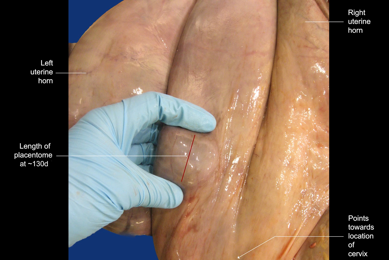
The image below shows the general area in which placentomes are palpated. In essence, one only uses the placentomes close to the cervix to make estimations of the size of these structures. If one palpates more cranial than this, it is likely that the duration of the pregnancy will be over estimated because placentomes generally become larger as one's hand moves cranially over the “pregnant” horn.


Image size: 3660x 2112px

Although the third image shows a placentome conveniently placed between thumb and forefinger for a measurement, this is not always possible in practice. More often, to gain an impression of their average size, several placentomes are manipulated between one's fingers while pressing down within the rectum.


Image size: 3152x 2112px

The image below shows how placentomes grow during pregnancy. Note that their growth is almost logarithmic in the last third of gestation.  The benchmarks discussed in the paragraph below are colored green in the image.


Image size: 1492x956px


Every operator will have her or his preferred method of remembering the size of placentomes at any given stage of pregnancy. The author provides the following guidelines for consideration. Again, these guidelines are used for palpation in the absence of ultrasonography. Measurements of structures using ultrasonography are highly accurate but when one takes tissue compression (during transrectal palpation) into account, mental impressions are not the same as as ultrasonographic measurements.

At 75 days, placentomes usually become discernible for the first time.

At 90 days (3 months) placentomes measure about 1 x 1.5 cm in size. In the author's rather unconventional mind, it is easily remembered because 1.5 is half of 3 but admittedly, this is barely rational.

At 120 days (4 months) placentomes measure approximately 2.5 x 1.5 cm in size. Added together, 2.5 and 1.5 equal 4 ; easy to memorize.

At 150 days (5 months) placentomes measure approximately 2 x 3 cm in size. Added together, 2 and 3 equal 5 ; easy to memorize.

After 5 months, the remarkable growth rate of placentomes become noticeable.

The dimensions of placentomes at 5 months of gestation are particularly useful to memorize because they are about that size in beef cattle during routine fall pregnancy diagnosis. Simply by spreading one's fingers slightly and applying pressure on the dorsal  surface of the uterus, then “raking’ in a cranial to caudal motion, a five month old pregnancy can be diagnosed easily. This procedure is so rapid and simple that hundreds beef cows can be examined in a few hours

At 6 months of gestation, the placentomes are approximately 6 cm long; again this is easy to memorize.

At term, the rapid growth of placentomes has increased their average length to approximately13 cm. Again, in the author's unconventional mind, this is remembered as “unlucky 13 at term".

Again, these benchmarks are 75 days, 3 months, 4 months, 5 months, 6 months and Term. Other durations are extrapolated between these benchmarks.
_____________________________

In the event that a fetus is aborted and one has to determine at what stage of gestation that occurred, many different guidelines are provided in the literature, some bordering on the ludicrous (small mouse, large rat without a tail etc). Instead, the following simple guidelines are offered. Again, one uses the dimensions of one's hand to make these determinations. A discussion follows the image.


Image size: 3046x2437px

The crown-rump length (CRL) of a two month old fetus extends across the palm of the author's hand as shown. At four months, the CRL extends between the tip of the author's middle finger and his wrist. Placing both hands, tip-to-tip as shown, covers the CRL of a six month old fetus. Finally, beyond the combined size of one's hands, the CRL increases to 80 cm at 8 months. Interestingly, these measurements apply to all ruminants and even horses. Incidentally, the CRL of an aborted 10 month old equine fetus is approximately 100cm.